

Abstract
Background
Alzheimer’s disease (AD) is characterized by amyloid plaques, tau tangles, and neuronal loss. Progranulin (PGRN) is a secreted immune regulator, lysosomal chaperone, and neuronal survival factor. Genetic polymorphisms that reduce PGRN levels are associated with an increased risk for AD and other neurodegenerative disorders. The receptor sortilin binds and targets PGRN for lysosomal degradation, resulting in a reduction of extracellular PGRN. AL101 (GSK4527226) is a monoclonal antibody that binds to the sortilin receptor and is being developed as a potential PGRN-elevating therapy for AD.
Methods
Cell-based in vitro studies examined the interaction of AL101 with sortilin and its effect on PGRN levels. In vivo studies evaluated the safety, pharmacokinetics (PK), and pharmacodynamics (PD) of AL101 in rats and nonhuman primates. A phase 1 study in healthy volunteers assessed PK, safety, tolerability, and PD biomarkers after intravenous or subcutaneous dosing of AL101.
Results
Cell-based assays showed that AL101 increased PGRN levels by decreasing cell surface sortilin levels and partially blocking the sortilin-PGRN interaction. Preclinical studies in rats and monkeys demonstrated that AL101 decreased cell surface sortilin levels on white blood cells and increased PGRN levels by up to 2-fold in cerebrospinal fluid (CSF) and up to 4-fold in blood. In the phase 1 study in healthy volunteers, both single and multiple doses of AL101 led to significant increases in plasma and CSF PGRN levels, providing additional support for its potential as a PGRN-elevating therapy.
Limitations
The first-in-human dose-finding study was aimed at investigating the safety and tolerability of AL101 and was not sufficiently powered to detect changes in exploratory outcomes, such as neurodegeneration biomarkers. Clinical studies are needed to evaluate AL101 in AD patients.
Conclusions
AL101 was shown to bind sortilin and decrease cell surface sortilin levels, leading to consistent elevations of PGRN across in vitro, preclinical, and human studies. These results support continued development of AL101 and its investigation as a potential treatment for AD and other neurodegenerative conditions where PGRN could play a role.
Trial registration
Clinicaltrials.gov, NCT04111666. Registered on October 1, 2019. https://clinicaltrials.gov/ct2/show/NCT04111666.
Background
Alzheimer’s disease (AD) is a progressive neurodegenerative disorder that primarily affects individuals aged 65 years and older. Approximately 6.7 million Americans live with AD [1]. The hallmark neuropathological features of AD include the accumulation of extracellular amyloid plaques and intracellular neurofibrillary tangles, which are initially seen in the medial temporal structures, leading to hippocampal atrophy and episodic memory impairment [2]. As the disease progresses, it affects other cognitive domains, eventually resulting in loss of independence in daily functioning [3, 4]. Risk factors for AD include age, comorbid medical conditions (e.g., cardiovascular disease, recurrent depression, history of head trauma), and genetic determinants [5,6,7]. Current symptomatic therapies for AD, such as acetylcholinesterase inhibitors and N-methyl-D-aspartate receptor antagonists, provide modest benefits to cognitive and behavioral parameters but do not affect disease progression [8,9,10,11]. Recently, monoclonal antibody treatments targeting amyloid beta have been approved for the treatment of mild cognitive impairment (MCI) or mild dementia due to AD. However, the long-term effects of these treatments remain to be characterized, and the impact of safety concerns observed about amyloid-related imaging abnormalities, such as edema and microhemorrhages within the brain are not fully understood [12,13,14,15]. Novel therapies with different mechanisms of action that can improve efficacy and safety are urgently needed, either as monotherapies or in combination with anti-amyloid therapeutics [16].
While there is no single underlying cause of AD, recent advances have identified several underlying genetic risk factors. Many of the genes that have been implicated in AD underpin immune response regulation, amyloid precursor protein and tau processing, lipid biology, and lysosomal function/endocytosis [17,18,19]. Among the recently discovered AD risk genes is GRN [20,21,22,23,24,25,26], which encodes progranulin (PGRN), a secreted protein that acts as a lysosomal chaperone, neurotrophic factor, and regulator of immune cells [27,28,29,30,31,32,33,34,35]. PGRN is widely expressed in neurons and microglia and plays a critical role in maintaining microglial homeostasis, neuronal survival, and neurite outgrowth [29,30,31,32, 34]. Notably, GRN polymorphisms associated with reduced PGRN levels modify the risk for AD [36]. For example, the rs5848 T-allele has been found to increase the risk of AD and is associated with decreased levels of serum and CSF PGRN [20, 26, 37, 38]. Additionally, a study of AD patients found the single nucleotide polymorphism (SNP) rs5848 to be consistently associated with decreased GRN mRNA in the parietal lobe and also found disease onset to be earlier in a subset of patients carrying the rs9897526A variant [39]. GRN polymorphisms in patients clinically diagnosed with AD suggests that such mutations may be a risk or that they may play a disease-modifying role, influencing the age of onset by promoting a proinflammatory state that contributes to neuronal death in AD [22, 23, 39, 40].
In addition to the linkage between GRN SNPs and AD, homozygous loss-of-function GRN mutations that result in a complete loss of PGRN cause neuronal ceroid lipofuscinosis, a rare lysosomal storage disorder, and heterozygous loss-of-function GRN mutations, which reduce PGRN levels by ~ 50%, can cause frontotemporal dementia (FTD) [41,42,43,44,45,46,47]. It is notable that there are over 100 GRN-heterozygous, loss-of-function mutations that have been identified in clinical populations, potentially leading to a variety of neurodegenerative conditions due to lowered PGRN levels [48, 49]. For example, among patients with amyotrophic lateral sclerosis (ALS), common variants in GRN have been associated with earlier age of ALS onset and shorter survival time [50]. Parkinson’s disease has also been associated with risk variants related to GRN, and plasma PGRN levels in patients with Parkinson’s disease are decreased relative to healthy controls and negatively correlated with disease severity [33, 51, 52].
Given the strong link between GRN loss-of-function mutations and neurodegeneration [24, 53], increasing PGRN levels could potentially be an effective therapy for multiple disorders [36, 54,55,56,57]. Alterations in cerebrospinal fluid (CSF) PGRN levels have been observed in neurodegenerative conditions such as AD, with reduced levels during symptom onset and elevated levels throughout disease progression, potentially resulting from an insufficient microglial response to amyloid deposition and pathology [58]. Elevating PGRN early in the disease course has the potential to enhance protective microglia and lysosomal functionality, improving neuronal survival and thereby providing broad protection in AD.
PGRN is endocytosed and negatively regulated by sortilin, which is a sorting and scavenging receptor [59, 60]. Sortilin operates on the cell surface and within the cell’s endoplasmic reticulum-Golgi apparatus to promote lysosomal degradation of PGRN [59, 60]. Blocking the sortilin-PGRN interaction reduces PGRN degradation, resulting in increased levels of secreted PGRN in the central nervous system (CNS) [61]. In animal models, suppression of SORT1 expression has been associated with increased PGRN levels, decreased microglial activation, and ameliorated inflammation [62]. Thus, reducing sortilin levels and its interaction with PGRN could be an effective strategy for elevating PGRN and altering disease progression [54, 56, 60, 63].
AL101 (GSK4527226) is a monoclonal antibody therapy in development for AD that targets sortilin to elevate functional PGRN levels in the CNS and potentially alter disease progression. In this study, we describe the target engagement and mechanism of action of AL101 in cell-based studies and evaluate the safety, pharmacokinetics (PK), and pharmacodynamics (PD) of AL101 in rats and cynomolgus monkeys. Furthermore, we evaluate the safety profile of AL101 and demonstrate its target engagement in a first-in-human (FIH) study.
Methods
In vitro characterization of AL101
AL101 is a human monoclonal immunoglobulin G (IgG) G1m17,1 kappa antibody generated against the human sortilin receptor. AL101 was generated and affinity-matured in collaboration with Adimab (Lebanon, New Hampshire, USA). Three-point mutations, L234L235 to A234A235 and P331 to S331 (Kabat numbering system; L246L270 and P343 in sequential numbering), were made to the heavy chain to minimize effector functions such as Fc gamma receptor binding, complement activation, and antibody-dependent cell-mediated cytotoxicity [64, 65]. The three-point mutations are intended to improve the overall safety of AL101 for chronic administration in humans.
In vitro characterization of AL101 was performed as previously described [61] with slight modifications. Briefly, AL101 binding to the sortilin receptor was assessed on human sortilin stably expressed in human embryonic kidney 293 (HEK293T) cells, and AL101 antibody blocking of PGRN binding to sortilin was performed via a cell-based competition assay [61] using recombinant PGRN. To assess the effect of AL101 on PGRN levels, surface sortilin and the levels of secreted PGRN were measured in human U251 cells plated at 15,000 cells per well 24 h prior to incubation with AL101 or isotype control. U251 cells were lifted using Cell Stripper (#25-056-CI), washed in 2% fetal bovine serum, and incubated with unlabeled, non-competing S2-11 anti-sortilin for 1 h on ice. Detection was with 5-µg/mL phycoerythrin (PE)-conjugated goat anti-human antibody (Southern Biotech #2040-09) for 30 min on ice using the BD fluorescence-activated cell sorting (FACS) Canto II for analysis of mean fluorescence intensity of PE signal. Secreted PGRN was assessed in the culture media using a commercial enzyme-linked immunosorbent assay (ELISA) kit (R&D Cat. #DY2420), plates were read on a Biotek Synergy HT Multi-Detection Microplate Reader, and data were graphed and analyzed as previously described using GraphPad Prism [61].
PK and PD of AL101 in rats
Male and female RccHan:WIST rats (99 animals/sex), 10 to 11 weeks of age, were randomly assigned to receive once weekly intravenous (IV) injections (for a total of 5 injections) of 0- (vehicle), 40-, 100-, or 300-mg/kg AL101. Noncompartmental analysis (NCA) methods were applied to the composite serum AL101 concentration for PK analysis.
Blood samples for PD (serum PGRN and white blood cell [WBC] sortilin levels) were collected from 3 animals/sex/dose/group at pre dose and approximately 0.5, 4, 24, 72, 168, and 336 h post dose after the first (Day 1) and fifth (Day 29) dose. CSF can only be collected from rats at termination. Terminal CSF samples for PK and PD (PGRN) were collected from 3 animals/sex/group on Day 29 at approximately 24, 168, and 336 h post dose. For WBC processing, 1 mL of ammonium-chloride-potassium (ACK) lysis buffer was added to 200-µL whole blood, mixed, and incubated at room temperature for approximately 5 min, followed by centrifugation at 500 g for 10 min. The cell pellets were frozen at − 80 °C until used. For sortilin concentrations, high-binding 96-well ELISA plates were coated with an anti-sortilin antibody (1 µg/mL) in phosphate-buffered saline (PBS) at 4 °C overnight. Diluted WBC lysate samples were added to the wells for 1 h at room temperature. Wells were then incubated with goat anti-mouse sortilin-biotinylated secondary antibody (0.1 µg/mL; R&D Systems) for 1 h followed by streptavidin-horseradish peroxidase (HRP) (R&D Systems) for 30 min. Pierce 1-step ultra tetramethylbenzidine (TMB) was used for visualization and the reaction was stopped with 2 N sulfuric acid.
Concentrations of PGRN were measured by ELISA following manufacturer’s instructions (AdipoGen Life Sciences; Cat. #AG-45A-0043YEK-KI01; Lot #K5211908 or R&D Systems; Cat. #DY2420; Lot #P325650). Optical densities were measured at 450 nm using a Molecular Devices M5 or a Biotek Synergy H1 microplate reader. Data were analyzed using SoftMaxPro, Excel, and GraphPad Prism 7.0.
PK and PD of AL101 in cynomolgus monkeys
In this GLP toxicology study, 36 cynomolgus monkeys (n = 18/sex), 32 to 57 months of age, were administered doses of 0- (vehicle), 20-, 60-, or 200-mg/kg AL101 once weekly via IV injection (for a total of 5 injections). During the dosing phase, blood samples were collected at pre dose and approximately 10, 48, and 96 h post dose on all 5 dosing days, at approximately 2 and 144 h post dose on Days 1 and 22, and at pre dose and 2 h post dose on Day 29. During the recovery phase, blood samples were collected from the vehicle and 200-mg/kg AL101 groups at 96, 168, 336, 504, 672, 840, and 1008 h post last dose. CSF samples were collected from anesthetized animals via the cisterna magna once during the predose phase, approximately 2 h post dose after the first (Day 1) dose, and on days of scheduled sacrifice. Serum levels of AL101, WBC sortilin, serum PGRN, and CSF PGRN were assayed as described above. Clinical observations were performed at predose baseline as well as before and after drug administration on all dosing days and on scheduled days of sacrifice.
FIH phase 1 study of AL101 in healthy volunteers
Study design
This FIH phase 1 study was a randomized, double-blind, placebo-controlled trial designed to investigate the safety, tolerability, PK, and PD of single doses (SDs) and multiple doses (MDs) of AL101 administered by IV every 4 weeks (q4w) (Fig. 1). AL101 dose levels evaluated were 6, 15, 30, and 60 mg/kg. The study also incorporated SD (150 mg and 600 mg) and MD subcutaneous (SC) doses (300 mg every 2 weeks [q2w] for a total of 7 doses) of AL101 to further evaluate bioavailability using a different route of administration.

Study design. This was a FIH phase 1 study designed to investigate the safety, tolerability, PK, and PD of SDs and MDs of AL101 administered by IV and SC routes. Bioavailability of AL101 was evaluated for SC administration. The study had 2 parts. The first part was a randomized, double-blind, placebo-controlled, SAD study of AL101 vs. placebo administered IV. The second part studied SC dosing (SD and MD) and MD IV administration. Although the study was outlined as a 2-part study, the data are presented by overall dosing scheme, by SD and MD cohorts separately. Serum samples for pharmacokinetic analyses and plasma samples for pharmacodynamic analyses were collected at each visit. CSF samples were collected at baseline and at 2 time points post dose; the schedule of post dose CSF sampling varied for different subgroups. a Treatment day. b Postdose CSF sample collection. Note that the schedule of postdose CSF sampling varied for different subgroups in the SAD IV and 600-mg SC cohorts. CSF, cerebrospinal fluid; FIH, first-in-human; IV, intravenous; MD, multiple dose; PK, pharmacokinetics; PD, pharmacodynamics; q2w, every 2 weeks; q4w, every 4 weeks; SAD, single-ascending dose; SC, subcutaneous; SD, single dose
The dosing schemes in the FIH study were guided by results from the nonclinical, toxicological rat and cynomolgus monkey studies. Note that in the nonclinical toxicology studies, the lowest evaluated dose within each species was determined based on an approximation of the clinical dose, the mid and high doses were anticipated to be significant multiples of the clinical dose on a pharmacokinetic equivalent basis, and the high dose was also an approximation of the maximum feasible dose based upon dosing volume considerations. The toxicology studies revealed no AL101-related adverse events (AEs) up to the highest dose tested (i.e., 200 mg/kg and 300 mg/kg in cynomolgus monkeys and rats, respectively). SD, body-weight-based safety margins were based on the no observed adverse effect level (NOAEL) in cynomolgus monkeys because this was the more sensitive species based on body weight dosing. Specifically, a safety factor of 10 was applied to the NOAEL of 200 mg/kg, yielding a maximum recommended starting dose (MRSD) of 20 mg/kg. AL101 was expected to be well tolerated by healthy participants at a lower starting dose of 6 mg/kg IV, which was 30% of the MSRD (i.e., a safety margin of 33.3 to the cynomolgus monkey NOAEL). In cynomolgus monkeys, CSF PGRN was increased by all AL101 doses (see results below), so it was expected that PGRN increases in human CSF would be seen at the higher of the planned IV dose levels (15, 30, and 60 mg/kg). MD safety margins were based on the area under the concentration-time curve (AUC) from time 0 to the end of the dosing interval (AUC0 − tau) and steady-state maximum observed concentration (Cmax) at the NOAEL from a 26-week repeat-dose GLP toxicology study in rats (i.e., 300 mg/kg). The MD cohorts were added to the protocol to refine the understanding of the safety, PK, and PD profile of AL101 after clinical evaluation of the safety data from the SD cohorts.
Participants were randomized to AL101 or placebo in a ratio of 8:3 for IV and SC dosing. AL101 or placebo was administered as a single peripheral IV infusion over approximately 60 min, and SC administration was done with slow injection over the course of 15 min. Sentinel dosing occurred in the first participant of the 6-mg/kg IV cohort and 300-mg/kg SC cohort. Subsequent SD and MD SC and IV cohorts proceeded without sentinel dosing. For the SD IV cohorts, dose escalation decisions were made after all participants who were assigned to a given dose level were dosed and sufficient safety data were available, as defined by the dose-limiting adverse event (DLAE) assessment window. The DLAE window began at the start of infusion on day 1 and ended 12 days after the infusions. To proceed with dose escalation, a minimum of 6 participants were required to complete the DLAE window in a given SD IV cohort. The decision to escalate to the next dose level was based on the assessment of safety/tolerability, ECG, vital signs, and clinical laboratory data up to and including the most recently dosed cohort and any cumulative data.
Serum and plasma samples for PK and PD analyses were collected at prespecified time points (Fig. 1). The PK endpoints included serum and CSF concentrations of AL101 at various time points and standard PK parameters. The PD endpoints included the change in PGRN levels in plasma and CSF after dosing relative to baseline. ELISA PGRN levels have been shown to be highly correlated between serum and plasma [66], so the shift in matrix type between preclinical and clinical studies was not expected to impact this study’s findings. Safety endpoints included the incidence, nature, and severity of AEs, incidence of DLAEs, incidence of treatment discontinuation due to AEs, mean changes in clinical laboratory test results or vital signs from baseline over time, physical and neurologic abnormalities, and incidence of anti-drug antibodies (ADAs). An AE was considered a DLAE if it was assessed as related to study drug in participants who received AL101 and could include any of the following: any serious or ≥ Grade 3 AE with no other clearly attributable cause beyond study drug, or a ≥ Grade 2 infusion-related toxicity occurring within 24 h of the infusion. Decisions to escalate doses in the single-ascending dose (SAD) IV cohorts were made by a safety review committee (SRC) based on all available safety, tolerability, PK, and PD data from previously dosed participants. The DLAE window began at the start of infusion and ended 12 days later.
Participants
Males and females (who were using contraception and confirmed not pregnant) aged 18 to 65 years were included in the study. Participants also had to be in good physical health, be non- or light (< 5 cigarettes/week) smokers, abstain from alcohol consumption for 48 h prior to blood collections and clinical laboratory tests, and stop all over the counter or prescription medications within 14 or 30 days of study admission, respectively. Exceptions were made for use of acetaminophen/paracetamol, hormonal contraceptives, and medications for hypertension. Participants were excluded from the study if they met any of the following criteria, among others: known history of reactions to antibodies or fusion proteins; positive drug or alcohol results at screening and prior to first dose; history of alcohol or substance abuse within the past 2 years; history of seizures, major depression, schizophrenia, schizoaffective disorder, or bipolar disorder; history of cancer with exceptions; chronic kidney disease or impaired hepatic function; clinically significant cardiovascular disease or uncontrolled hypertension; or any serious medical condition or abnormality. Additionally, participants in the SC AL101 cohorts were excluded if they had tattoos, scars, or sunburn in any of the designated injection sites, or if they had a history or presence of a skin rash or disorder.
Bioanalytical procedures
Electrochemiluminescent assays (ECLAs) were used to quantify AL101 concentrations in CSF and serum samples. Recombinant human sortilin (R&D Systems) was diluted in coating buffer, refrigerated for up to 1 day, the plate was washed, and then all unabsorbed sites were blocked with blocking buffer. A 1:10 assay buffer was added followed by the addition of biotinylated anti-AL101 20D12, SULFO-TAG streptavidin, and finally a 1x read buffer T (Meso Scale Diagnostics), with washes and 30- to 60-minute incubation periods between each step. AL101 concentrations were determined on a standard curve plotting relative light units (RLUs) vs. concentration using a 4-parameter curve-fitting program with 1/y2 weighting.
PGRN levels in plasma and CSF were determined via ELISA following manufacturer’s instructions (AdipoGen Life Sciences, Cat. #AG-45A-0043YEK-KI01) using a minimum required dilution (MRD) of 1:40 for plasma and 1:4 for CSF and with recombinant human PGRN (R&D Systems, Cat. #2420-PG) or equivalent as a calibrator.
Statistical analysis
All statistical analyses for the preclinical studies were performed using GraphPad Prism. Statistical significance was determined using 2-way analysis of variance (ANOVA) for the in vivo animal studies.
All phase 1 statistical analyses were conducted using SAS Enterprise version 7.1 (SAS Institute). Summaries were presented by dose level and participants receiving placebo were combined into a single group per route of administration. Frequencies and percentages were presented for categorical variables, and descriptive statistics were summarized for continuous variables. Participant disposition and demographic information were summarized for the enrolled population. All safety analyses were conducted on the safety population, which included all enrolled participants who received at least 1 dose of AL101. No formal statistical inferences were made for safety parameters, and no imputation was used for missing data.
Mean serum and CSF concentrations of AL101 were calculated for each nominal time point. The individual serum concentration vs. time data for AL101 was used to derive the PK parameters by NCA methods using Phoenix WinNonlin Version 8 (Pharsight Corporation). Geometric mean and 95% confidence intervals were presented for PK parameters. Dose proportionality for AL101 was evaluated for Cmax, AUC0 − tlast, and AUC0 − inf. A power model was fitted to describe the relationship between 6 mg/kg and 30 mg/kg using a least-squares linear regression model. Dose proportionality was concluded if the 90% CI of the slope lay entirely within (0.6990, 1.3010). For PD, baseline was defined as the last non-missing assessment prior to the first drug administration. Percent change from baseline in plasma and CSF PGRN was derived by time point for each participant and then summarized as mean changes from baseline for each treatment and dose level.
Study approval
The study was conducted in accordance with the guidelines set forth in the International Council for Harmonisation of Technical Requirements for Pharmaceuticals for Human Use (ICH), the Declaration of Helsinki, the study protocol, and all national, state, and local laws or regulations. Informed consent was obtained from each participant prior to study entry and prior to any study-related procedures.
Results
AL101 blocks PGRN binding to sortilin and decreases cell surface sortilin levels in vitro
AL101 is a recombinant human anti-sortilin monoclonal IgG1 antibody that binds the beta propeller domain of sortilin, which is where the C-terminus region of PGRN binds [60]. The sortilin binding affinity of AL101 was assessed by FACS using an HEK293T line with stable overexpression of human sortilin. We found that AL101 exhibited high-affinity binding for sortilin, with a mean half-maximal effective concentration (EC50) of 1.614 nM (Fig. 2A). To investigate the blocking effect of AL101 on PGRN binding to sortilin, a cell-based competition assay was conducted using biotinylated PGRN and the sortilin-overexpressing HEK293T cells. A dose-dependent partial inhibition of biotinylated PGRN binding to sortilin in the presence of AL101 was observed (Fig. 2B), with a range of 35–64% maximal blocking at 150 nM, the highest concentration assessed (average blocking maximum observed at 49% ± 12.9%). Next, we assessed the impact of AL101 on endogenous sortilin levels in human U251 cells and found that AL101 dose-dependently reduced cell surface levels of sortilin by 76%. In contrast, cells treated with an isotype control antibody showed no significant change in surface sortilin expression (Fig. 2C). The mean EC50 for the decrease in cell surface sortilin was determined to be 0.186 nM. In parallel, extracellular PGRN in the cell culture media supernatants was measured by ELISA, and AL101 treatment dose-dependently increased PGRN levels, with an average 55% elevation compared to treatment with an isotype control (Fig. 2D). In these in vitro experiments, AL101 reduced the cell surface expression of sortilin, partially blocked the binding of PGRN to sortilin, and resulted in a concomitant increase in extracellular PGRN levels.
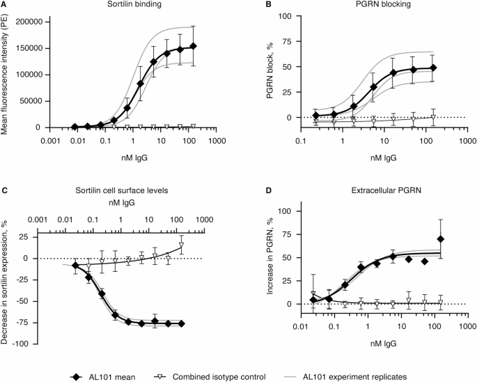
AL101 in vitro binding characterization. (A) Binding curve of human sortilin-overexpressing HEK293T cells by AL101 and an isotype control human IgG. (B) Percentage of dose-dependent blocking of PGRN binding to sortilin by AL101 and isotype control human IgG. Percent block calculated by normalizing to no antibody treatment conditions. (A, B) Combined average of n = 3 experiments with AL101 represented by the black line with diamond points; individual experiments shown with gray points and curves; combined average of n = 2 to 3 experiments with isotype control represented by open triangles. (C) Dose-dependent decrease in surface sortilin levels on U251 cells by AL101 and isotype control human IgG. (D) Increased extracellular PGRN by AL101 treatment relative to isotype control–treated cells. (C, D) The mean ± standard deviation of n = 2 to 3 experiments with AL101 is represented by the black line with diamonds; individual experiments are represented by gray curves; the mean ± standard deviation of n = 2 to 3 experiments with human IgG isotype control is represented by open triangles. HEK293T, human embryonic kidney 293 cells; IgG, immunoglobulin G; PE, phycoerythrin; PGRN, progranulin
Weekly AL101 administration in rats decreases sortilin levels in WBCs and increases PGRN levels in serum and CSF
The concentration-time profiles of AL101 in serum indicated systemic exposure to the drug throughout the intended treatment duration (Supplementary Fig. S1). Cmax and area under the concentration-time curve between 0 and 168 h after dosing (AUC0 − 168) of AL101 increased in a generally dose-proportional manner from 40 to 300 mg/kg/dose (Supplementary Table S1). Exposure was generally higher on Day 29 compared to Day 1, indicating some accumulation of AL101 following multiple weekly doses (Supplementary Table S1).
Safety assessments were conducted throughout the 4-week GLP toxicology study. There were no AL101-related clinical observations, changes in body weight or food consumption, or effects on hematology, coagulation, clinical chemistry, or urinalysis. There were no occurrences of AL101-related dermal observations or AL101-related elicited behavior, open-field response, or neurobehavioral differences. Additionally, there were no occurrences of AL101-related mortality, and no AL101-related effect on organ weights or microscopic or macroscopic observations upon terminal evaluation. Overall, dose levels up to the highest tested dose of 300 mg/kg were well tolerated without adverse findings, establishing the NOAEL as 300 mg/kg/dose.
Within 24 h of the first AL101 dose, the levels of sortilin in WBCs significantly decreased in all AL101-treated groups compared to the control group (P < 0.05, 2-way ANOVA; Fig. 3A). This decrease in sortilin levels was sustained throughout the study period, up to 336 h after the last dose (P < 0.05, 2-way ANOVA; Fig. 3A). There were no significant differences observed between the different AL101-treated groups.

Weekly dosing of AL101 in rats results in a chronic reduction of WBC cell surface sortilin levels and a parallel elevation of PGRN in serum and CSF. (A-C) Concentration-time profiles of sortilin cell surface levels in rat WBCs (A) and PGRN in rat serum (B) and CSF (C). All data represent the mean ± SEM. Arrows indicate the timing of injections. For WBC sortilin, n = 3 to 6/group/time point; for serum PGRN, n = 5 to 6/group/time point; for CSF PGRN, n = 4 to 17/group/time point. Only the placebo and 300-mg/kg dose groups were included in the 6-week recovery phase. CSF, cerebrospinal fluid; PGRN, progranulin; SEM, standard error of the mean; WBC, white blood cell
Furthermore, serum levels of PGRN were significantly increased in AL101-treated animals compared to the control group as early as 4 h after the first dose (P < 0.05, 2-way ANOVA; Fig. 3B). The increase in serum PGRN levels continued in all AL101-treated groups up to 24 h after the initial dose and remained at similar levels for up to 336 h after the last dose (Fig. 3B). The elevation of serum PGRN levels was consistent across different dose groups, indicating that even at the lowest AL101 dose of 40 mg/kg, maximum elevation of PGRN levels was achieved in the peripheral circulation.
Similar to serum, there was a significant increase in CSF PGRN levels in all rats treated with AL101 compared to the control group (P < 0.05, 2-way ANOVA; Fig. 3C). CSF PGRN levels remained significantly elevated in the 300-mg/kg AL101 group compared to the control group for up to 6 weeks after the last dose (Fig. 3C). For each timepoint and dose group, a subset of rats was sampled for WBC cell surface sortilin levels and serum PGRN concentrations. Thus, normalization of sortilin or PGRN concentrations relative to predose baseline was not feasible for individual rats and only average concentration of sortilin and serum PGRN levels were reported. Further, CSF was only collected once in each rat at termination, thus no predose CSF PGRN concentrations were reported. Amongst the AL101 dosed groups, there were no significant differences in CSF PGRN levels between the 40-, 100-, and 300-mg/kg dose groups, except at 720 h post dose where a significant difference was observed between the 40- and 300-mg/kg groups. These results indicate that weekly administration of AL101 in rats led to a decrease in sortilin levels in WBCs and an increase in PGRN levels in both serum and CSF.
Repeat dosing of AL101 in cynomolgus monkeys decreases cell surface sortilin levels in WBCs and increases PGRN levels in serum and CSF
The serum concentration-time profiles demonstrated that AL101 was detectable in the monkeys throughout the dosing and recovery periods (Supplementary Fig. S2; Supplementary Table S2). AL101 exposure increased with higher doses and showed a generally dose-proportional relationship (Supplementary Table S2). Accumulation of AL101 was observed in monkeys after weekly dosing, with higher exposure levels on Days 8, 15, 22, and 29 compared to Day 1 (Supplementary Table S2). An immune response against AL101, as indicated by reduced exposure consistent with an ADA response, was observed in 1 animal in the 200-mg/kg dose group starting from Day 15. However, no evidence of an ADA response was observed in other animals.
During the dosing and recovery phases, no AL101-related effect was identified in clinical observations or in hematology, coagulation, clinical chemistry, or urinalysis test results. Additionally, no AL101-related mortality occurred during the dosing or recovery phase, and there was no AL101-related effect on organ weights or anatomic pathology. Overall, dose levels up to the highest tested dose of 200 mg/kg were well tolerated without adverse findings, establishing the NOAEL as 200 mg/kg/dose.
AL101 treatment led to significant reductions in cell surface sortilin levels in WBCs compared to vehicle control, starting at 96 h after the first dose (Fig. 4A; P = 0.0158, P = 0.0077, and P = 0.0009, respectively, for the 20-, 60-, and 200-mg/kg dose groups vs. vehicle control, 2-way ANOVA). Sortilin levels in all 3 AL101 treatment groups remained consistently decreased vs. placebo throughout the study period (Fig. 4A). There was no significant difference between the AL101 treatment groups. Serum PGRN concentrations were significantly increased in the AL101-treated groups compared to vehicle control, starting at 48 h after the first dose (P = 0.033, P = 0.001, and P = 0.0008, respectively, for the 20-, 60-, and 200-mg/kg dose groups vs. vehicle control, 2-way ANOVA) and lasting up to 144 h post dose (Fig. 4B). The increase in serum PGRN levels was 3- to 4-fold higher than baseline and vehicle control values. This elevation in serum PGRN levels was consistent across different dose groups, indicating that even at the lowest AL101 dose of 20 mg/kg, the maximum elevation of PGRN levels in the periphery was reached. PGRN levels remained elevated at least 3-fold above baseline throughout the study duration and up to 6 weeks after the last dose in 3 out of 4 animals in the 200-mg/kg group during the recovery phase.

Multiple dosing of AL101 in cynomolgus monkeys decreases WBC cell surface sortilin levels and increases PGRN levels. (A-C) Concentration-time profiles of sortilin expression in monkey WBCs (A) and PGRN in monkey serum (B) and CSF (C). Arrows indicate the timing of injections. Only the vehicle control and 200-mg/kg groups were included in the 6-week recovery phase. All data were normalized relative to predose baseline values (dashed lines) for each individual animal and are represented as the mean ± SEM. n = 4 to 10/group/time point. CSF, cerebrospinal fluid; PGRN, progranulin; SEM, standard error of the mean; WBC, white blood cell
Similar to serum, a significant increase in CSF PGRN levels was observed in all AL101 dose groups compared to vehicle control at all measured postdose time points starting at 48 h after the first dose (P = 0.019, P = 0.015, and P = 0.0004, respectively, for the 20-, 60-, and 200-mg/kg dose groups vs. control, 2-way ANOVA) (Fig. 4C). CSF PGRN levels remained elevated at least 2-fold above baseline and lasted up to 6 weeks after the last dose in 3 out of 4 animals in the 200-mg/kg group during the recovery phase. These results thus demonstrated that AL101 can not only increase PGRN in the CNS in nonhuman primates, but that the increase in CSF PGRN is a relatively durable effect.
Evaluation of SD or MD SC or IV administration of AL101 in healthy volunteers
Participants
The study enrolled a total of 61 participants in the SD cohorts, with 49 participants receiving AL101 and 12 participants receiving placebo (Supplementary Table S3). Demographic and baseline characteristics for participants treated with AL101 are presented in Supplementary Table S4. In summary, the demographic and baseline characteristics were balanced across treatment groups.
Among the participants who received SD AL101 administration, 48 participants (98.0%) completed the treatment, and 42 participants (85.7%) completed the study. The reasons for not completing the study included 1 participant (2.0%) who withdrew due to a treatment-emergent adverse event (TEAE; infusion-related reaction), 2 participants (4.1%) who discontinued based on physician decision, and 4 participants (8.2%) who withdrew from the study. In the SD placebo cohort, all participants completed the treatment, and 10 participants (83.3%) completed the study. Two participants (16.7%) in the SD placebo group withdrew from the study.
For the MD cohorts, a total of 27 participants were enrolled, with 24 participants receiving MD AL101 administration and 3 participants receiving placebo (Supplementary Table S3). Most participants receiving MD AL101 administration (79.2%) or placebo (66.7%) completed the study (Supplementary Table S3). Reasons for study discontinuation among participants receiving AL101 included 1 (4.2%) TEAE (infusion-related reaction) and being lost to follow-up (n = 4, 16.7%). One participant (33.3%) receiving placebo withdrew from the study.
Safety and tolerability
The SD cohorts included 46 participants (AL101, n = 34; placebo, n = 12) enrolled in the SD IV cohorts as well as 15 participants (600-mg AL101, n = 9; 150-mg AL101, n = 6) in the SD SC cohorts. Safety and tolerability results are presented in Table 1. Overall, 33 participants (67.4%) in the SD AL101 cohorts reported 64 TEAEs, and 7 participants (58.3%) in the SD placebo group reported 11 TEAEs. The most common TEAE among the AL101 treatment groups was headache, reported by 13 participants (26.5%). One participant in the 60-mg/kg SD IV group was withdrawn from the study drug due to an infusion-related reaction related to AL101. Another participant in the SD pooled placebo group had an isolated elevated alanine transaminase (ALT) level considered related to the study drug. No other clinically significant trends were observed in laboratory test results (hematology, serum, chemistry, or urinalysis), vital sign measurements, 12-lead electrocardiogram (ECG) results, neurological examinations, or physical examinations. No DLAEs were reported by the investigator for the SAD IV cohorts, but the sponsor determined that 1 event of infusion-related reaction in the 60-mg/kg SD IV group met the criteria of a DLAE. In the SD cohorts, 10 participants (21.3%) had treatment-emergent positive ADA (TEPADA) at any time during the study, including 1 participant who received placebo.
The MD cohorts included 11 participants receiving MD IV AL101 (30-mg/kg AL101, n = 11; placebo, n = 3) and 13 participants receiving 300-mg MD SC AL101. Among the MD AL101 groups, 16 participants (66.7%) reported 58 TEAEs, with headache being the most common (n = 7, 29.2%), followed by injection site erythema (n = 5, 20.8%) (Table 1). Three DLAEs occurred. In the 300-mg MD SC AL101 group, 1 serious AE (SAE) of myocardial infarction was reported in a participant with pre-existing atherosclerotic heart disease, which was deemed not related to the study drug. This participant also reported an event of severe acute respiratory syndrome coronavirus 2 (SARS-CoV-2) infection, which was mild in intensity and unrelated to the study treatment but considered a DLAE. Of the participants who received 30-mg/kg MD IV AL101, 1 participant had a nonserious TEAE of moderate infusion-related reaction leading to early discontinuation from the study; this event was considered related to study drug and was classified as a DLAE. Three participants in the MD cohorts (13.6%) had TEPADA results.
AL101 was distributed peripherally and centrally, as evidenced by serum and CSF AL101 concentrations
After a single IV infusion of AL101 at doses ranging from 6 to 60 mg/kg in healthy volunteers, the total exposure (area under the curve; AUC) and peak concentrations (Cmax) of AL101 in serum increased along with the dose (Fig. 5A; Supplementary Table S5). The increase in Cmax was dose proportional, while the increase in AUC was more than dose proportional based on statistical assessment of dose proportionality using a power model. The mean terminal elimination half-life (t1/2) of AL101 appeared to increase with increasing dose, ranging from 81.4 to 288 h (Supplementary Table S5). Total body clearance of AL101 decreased with increasing dose (Supplementary Table S5). Measurable concentrations of AL101 were detected for at least 43 days in the IV groups that received doses of at least 15 mg/kg (Fig. 5A).

Concentration-time profiles of AL101 from healthy volunteers. (A-B) Serum AL101 concentrations from participants in the SD (A) and MD (B) cohorts. Color-coded arrows indicate the timing of injections per group (B). (C) CSF AL101 concentrations from all cohorts plotted as a function of time. All data represent the mean ± standard deviation. For serum AL101, n = 2 to 13/group/time point, and for CSF AL101, n = 1 to 10/group/time point. CSF, cerebrospinal fluid; IV, intravenous; MD, multiple dose; SC, subcutaneous; SD, single dose
The mean total and peak exposures (area under the concentration-time curve from time 0 to infinity [AUC0-inf] and Cmax, respectively) of AL101 in serum appeared to increase in a greater than proportional manner with increasing dose, suggesting possible target-mediated drug disposition (Fig. 5A; Supplementary Table S5). The apparent elimination half-life and clearance of AL101 increased and decreased, respectively, with increasing dose in the SC groups (Supplementary Table S5). Measurable serum concentrations of AL101 were observed until Day 13 and Day 30 after administration of 150- and 600-mg doses, respectively.
After multiple IV or SC doses of AL101, the systemic exposure increased after the last administration, with little accumulation (Fig. 5C; Supplementary Table S5). The time to reach the maximum observed concentration (Tmax) was around 1 h after IV administration and around 70 to 72 h after SC administration. Serum concentrations of AL101 declined with mean elimination half-lives of 77 h and 286 h for IV and SC administration, respectively (Supplementary Table S5). Measurable serum levels of AL101 were observed until the last sampling time points for both MD IV and SC groups (Fig. 5B).
In terms of CSF concentrations, after a single IV infusion of 6 mg/kg or a single SC injection of 150 mg of AL101, the CSF concentrations were below the quantification limit (less than 7.81 ng/mL). CSF concentrations of AL101 appeared to increase in a dose-dependent manner in the IV groups receiving doses from 15 to 60 mg/kg (Fig. 5C). Quantifiable CSF levels of AL101 were observed at all planned collection days in the 30-mg/kg MD IV and 300-mg SD SC groups (Fig. 5C). The partition coefficient of AL101 was calculated as the ratio of CSF concentration to serum concentration for each matching timepoint. Following a single IV infusion of AL101 from 6 mg/kg to 60 mg/kg, the mean partition coefficient ranged from 0.00 to 0.00683 across the entire profile, indicating that the movement of AL101 from serum to CSF was less than 1%. The observed partition coefficient is consistent with the typical blood-brain barrier penetrance that has been observed with other peripherally administered monoclonal antibodies [67].
Plasma and CSF PGRN levels are elevated by SD and MD AL101 administration
A descriptive summary of the mean percentage change from baseline over time in plasma concentrations of PGRN for the SD and MD cohorts is depicted in Fig. 6A-B. In the SD cohorts, there was a dose-dependent numerical increase in the percentage change from baseline in plasma PGRN levels following a single IV infusion of AL101 ranging from 6 to 60 mg/kg or a single SC injection of AL101 ranging from 150 to 600 mg (Fig. 6A). Statistical assessment of the maximum change from baseline in plasma PGRN after IV administration of AL101 demonstrated a significant increase in plasma PGRN with all AL101 dose levels vs. the pooled placebo group; adjusted P < 0.0001 for the least squares mean (LSM) difference between the placebo group minus the 6-mg/kg (LSM difference = − 176.15), 15-mg/kg (LSM difference = − 186.27), 30-mg/kg (LSM difference = − 219.09), and 60-mg/kg (LSM difference = − 245.44) dose groups. Mean plasma PGRN concentrations returned to baseline levels on Day 43, 57, 85, and 113 after IV infusion of AL101 at doses of 6, 15, 30, and 60 mg/kg, respectively.

SD and MD AL101 administration increases PGRN levels. (A-B) Mean (± standard deviation) percentage change from baseline of plasma PGRN vs. time for SD (A) and MD (B) cohorts. Color-coded arrows indicate dosing days for the IV and SC MD cohorts (B). (C) Mean (± standard deviation) percentage change from baseline in CSF PGRN concentrations vs. time for all cohorts. For plasma PGRN, n = 1 to 13/group/time point, and for CSF PGRN, n = 1 to 10/group/time point. For plasma PGRN, n < 3 for the MD IV placebo group at multiple time points; for CSF PGRN, n < 3 for the SD IV placebo (Day 57), 6-mg/kg SD IV AL101 (Day 25), 30-mg/kg SD IV AL101 (Day 85), 600-mg SD SC AL101 (Day 43), and MD IV placebo groups (Days 97 and 113). CSF, cerebrospinal fluid; IV, intravenous; MD, multiple dose; PGRN, progranulin; SC, subcutaneous; SD, single dose
In the MD cohorts, AL101 resulted in numerical increases in plasma PGRN levels relative to baseline. Additionally, the percentage change in plasma PGRN was higher in the MD IV AL101 group compared to the pooled MD IV placebo group (Fig. 6B). Qualitative review of the descriptive summary statistics suggested that the percentage change from baseline in plasma PGRN appeared to reach a plateau after the first dose, regardless of the route of administration, and remained consistent for the subsequent doses (Fig. 6B).
Following SD and MD IV administration of AL101, the percentage change in CSF PGRN levels was numerically higher compared to the respective pooled placebo cohorts (Fig. 6C). Specifically, the maximum change from baseline in CSF PGRN levels increased to 77% and 67% in participants who received 15-mg/kg (Dunnett’s test, adjusted P = 0.0001) or 30-mg/kg (Dunnett’s test, adjusted P = 0.0005) SD IV AL101, respectively, compared to the pooled SD IV placebo group on Day 25. On Day 43, the maximum change from baseline in CSF PGRN significantly increased from 49 to 59% in participants who received either 15-, 30-, or 60-mg/kg SD IV AL101 compared to the pooled SD IV placebo group (Dunnet’s test, adjusted P < 0.0001 for 15-, 30-, and 60-mg/kg SD IV vs. placebo). MD IV administration of AL101 at 30 mg/kg q4w led to a numerical increase in CSF PGRN by 85.8% (1.86-fold) and 97.7% (1.98-fold) from baseline at Day 97 and Day 113, respectively, and MD SC administration of AL101 at 300 mg q2w led to an increase in CSF PGRN by 36.2% (1.36-fold) and 25% (1.25-fold) from baseline at Day 92 and Day 97, respectively.
Discussion
Current therapies for neurodegenerative diseases are lacking either in disease modification or are mostly for symptom relief. Therapies for AD, such as donepezil and memantine, show modest and transient cognitive and behavioral benefits, but do not slow or halt the progression of the disease [8,9,10,11]. Monoclonal antibody treatments targeting amyloid-beta (Aβ) have recently gained approval due to their demonstrated ability to slow decline in cognitive function in patients with AD. However, these treatments have also raised safety concerns such as amyloid-related imaging abnormalities (ARIA), that need to be carefully monitored [12,13,14,15]. Thus, there is still a significant unmet treatment need that will require the development of novel therapeutic agents.
PGRN has emerged as a potential therapeutic target in AD due to genetic, mechanistic, and therapeutic rationales [20, 35, 36, 42, 68]. GRN variants are among the genetic risk factors that have been shown to affect disease risk or progression in AD [20, 23, 24], as well as in other neurodegenerative diseases [42, 50, 51]. Consistent with this, in animal models, PGRN deficiency increased plaque load and impaired phagocytosis, whereas overexpression of PGRN reduced these deficits as well as amyloid burden [36, 68, 69]. Notably, GRN mutations have also been associated with Parkinson’s disease [51], and almost invariably lead to the development of FTD [33, 51], suggesting that targeting PGRN as a disease-modifying treatment might be a reasonable therapeutic approach across diseases, regardless of the underlying etiology. Therapeutics that elevate PGRN could have the potential to target not only amyloid and tau but also other proteinopathies, such as transactive response DNA-binding protein 43 (TDP-43), which is present in ~ 57% of AD subjects [70], and αsynuclein, which is present in ~ 30% of AD subjects [71]. Additionally, such therapies might enhance lysosomal function and promote neuronal survival [33, 72], thereby offering a more comprehensive clinical benefit compared to current therapies.
Here, we demonstrated the binding and target engagement of AL101 through in vitro cell-based assays and in vivo dose response studies in rodents, monkeys, and humans. AL101 bound to sortilin with high affinity, partially blocked the binding of PGRN to sortilin, and reduced cell surface sortilin levels, which resulted in elevated extracellular PGRN. The binding affinity of AL101 for sortilin is not expected to be tissue-dependent, so the kinetics of binding and internalization that were shown using WBCs from the periphery are expected to be consistent in blocking the sortilin-PGRN interaction in the CNS. In preclinical animal models, AL101 strongly decreased levels of WBC cell surface sortilin receptors in parallel to increasing PGRN levels in serum/plasma and CSF by blocking its uptake and degradation. PGRN can still enter the lysosomes, promote neuronal outgrowth, and reverse microglia pathology independent of its binding to sortilin [29, 32, 73, 74], suggesting that blocking and reducing sortilin does not impede the lysosomal, immune, and neurotropic effects of PGRN. An alternate mechanism for trafficking PGRN into the lysosome may occur through binding of prosaposin, which can bind mannose-6-phosphate receptor and low density lipoprotein receptor-related protein 1 and act independently of sortilin [74]. Additionally, studies utilizing sortilin knockout mice demonstrated that sortilin deficiency did not result in clinical or pathological manifestations of lysosomal storage disorders, that it was protective against age-dependent neurodegeneration and injury-induced cell death, and that it resulted in a 2.5-fold increase in total brain PGRN levels [60, 75, 76].
The target engagement of PGRN with AL101 administration was consistent across studies, with elevations detected in serum (rat and monkey), plasma (human), and CSF (rat, monkey, and human). In a prior study [61], we collected interstitial fluid (ISF) from the medial prefrontal cortex of mice that were administered AL101 or control antibody, and found that ISF PGRN levels in AL101-treated mice were ~ 3 times that of control-treated mice. Together, the statistically significant increase in CSF PGRN with AL101 vs. placebo in the rat, monkey, and human studies reported here and our earlier observation of increased ISF PGRN in mice, suggests that there is sufficient blood-brain barrier (BBB) penetrance of AL101 to increase PGRN levels in the brain. The therapeutic effect of these elevations in patients with AD is the subject of investigation in an ongoing phase 2 trial evaluating the safety and efficacy of AL101 in patients with early AD (PROGRESS-AD; NCT06079190). A robust PK/PD relationship was established and simulations from the PK/PD model informed the dose and regimen of AL101 treatment in the ongoing phase 2 study [77].
AL101 was generally well tolerated across the preclinical and clinical studies reported here. No toxicologically important effects of AL101 were observed in the rat and monkey GLP toxicology studies, supporting further development of AL101 in the FIH clinical study. There were no observed AL101-related AEs up to the highest doses tested in the preclinical toxicology studies. In the phase 1 dose-finding study, the rates of TEAEs were relatively comparable across AL101 dose groups, which were slightly higher than that observed in the corresponding placebo groups. Overall, there were 3 SAEs (1 each in the placebo, 60-mg/kg AL101 SD IV, and open-label 300-mg AL101 MD SC groups), 1 TEAE leading to discontinuation (30 mg/kg AL101 MD IV), and 3 DLAEs (1 each in the 60-mg/kg AL101 SD IV, 30-mg/kg AL101 MD IV, and open-label 300-mg MD SC groups).
In the CNS, PGRN is expressed on neurons and microglia and it plays a role in lysosomal health, neuronal survival, astrogliosis, and neuroinflammation [33]. Specifically, PGRN regulates lysosomal activity and inflammation in plaque-associated microglia, and it can promote microglial endocytosis of Aβ peptides and phagocytosis of Aβ plaques [78,79,80]. Deficits in PGRN can therefore contribute to the pathophysiology of AD via multiple mechanisms, including the aggregation of misfolded proteins, lysosomal dysfunction, complement activation, neuroinflammation, and astrogliosis, as well as aberrant microglial activation [33, 79, 81]. While the present set of experiments did not explore the effects of AL101 administration on markers of AD pathology, increased PGRN signaling is expected to improve the functionality of lysosomes to process lipids and misfolded proteins for degradation, have anti-inflammatory and neuroprotective effects, and promote Aβ clearance [81]. Given these hypothesized effects of increasing PGRN via sortilin blockade, AL101 may be a promising therapeutic strategy for other neurodegenerative diseases that have been associated with PGRN deficits, lysosomal dysfunction, or microglial risk factors. Various PGRN-elevating therapies, such as protein replacement (NCT05262023), an anti-sortilin monoclonal antibody (latozinemab, NCT04374136), and gene therapy (NCT04747431, NCT04408625), are currently being explored to restore PGRN levels in patients with FTD.
PGRN is composed of repeat domains that are proteolytically processed into granulins inside the lysosome. PGRN is critical for neuronal health, but the role of individual granulins is controversial. In preclinical models, granulins have been shown to increase inflammation, neurotoxicity, and lysosomal defects [82, 83]. Further, it has been shown that granulin cleavage product is accumulated in diseased brain regions from AD and FTLD patients [83]. Sort−/− mice have been shown to have an increase in the levels of full-length PGRN, with a corresponding reduction in granulin peptide levels from cortical lysates when compared to WT controls. This leads to an overall anti-inflammatory effect in which there is a significant decrease in the ratio between granulin peptides and full-length PGRN [84]. We would expect a similar reduction in granulins with AL101, leading to an overall similar anti-inflammatory effect.
AL101 may potentially have widespread effects on various cell types and processes, all of which remain to be elucidated. Here, we found that AL101 consistently decreased cell surface sortilin levels across species, and we observed strong evidence of target engagement of PGRN in the CNS. These findings do not preclude the possibility that decreasing cell surface sortilin levels might engage additional targets that mediate activity in inflammatory signaling pathways. For example, sortilin has a number of ligands other than PGRN that are involved in inflammatory processes and neuronal injury and repair, such as brain-derived neurotrophic factor (BDNF), amyloid precursor protein (APP), epidermal growth factor (EGFR), and neurotensin, among others [85,86,87,88]. Sortilin regulates neuronal viability and function, microglial activity, and cytokine exocytosis and signaling and it has also been implicated in AD [89,90,91]. Sortilin inhibition has been proposed to be a valuable therapeutic strategy for neurodegenerative diseases that include lysosomal dysfunction due to its potential to restore cellular recycling and maintenance pathways, disrupt downstream pathogenic cascades, and minimize cell death [89]. It is possible that the anticipated therapeutic effects of sortilin inhibition in AD might be at least partly mediated by blocking interactions with other known ligands in addition to increasing PGRN in the brain by blocking the sortilin-PGRN interaction. Further studies are needed to characterize the specific pathways that are impacted by AL101 administration and how such mechanistic effects will relate to clinical outcomes.
Limitations
The present set of studies has several limitations. First, the FIH study of AL101 consisted of a small number of healthy subjects, which may not be representative of the disease population of interest. Thus, it is not clear if the CSF PGRN elevations that were observed in healthy participants would be comparable to PGRN levels in patients with AD. Second, this study was aimed at investigating the safety and tolerability of AL101 across a range of doses in healthy volunteers, and it was not sufficiently powered to detect exploratory outcomes such as changes in neurodegeneration biomarkers. The effects of AL101-induced PGRN elevations on downstream targets that are known to play a role in AD pathology remain to be addressed. The study was also conducted in healthy volunteers, so it is unclear how the target engagement of PGRN by AL101 via sortilin blockade could potentially mitigate the development of AD pathology. Further investigation of the PD effects of AL101 in AD participants on key biomarkers of AD pathology, such as Aβ, tau, or neurofilament light, will be critical for understanding the disease-modifying potential of AL101. Third, AL101 has not been investigated in an established preclinical model of AD, so there is no precedent to extrapolate from regarding the potential slowing of decline in clinical (cognitive) measures. However, AL101 has been shown to increase PGRN levels in vivo and rescue a behavioral deficit in a mouse model of GRN haploinsufficiency [61]. Fourth, while the phase 1, dose-finding study included multiple dosing that extended for several months, the long-term safety of AL101 remains to be established.
AL101 is currently under investigation in a phase 2, proof-of-principle, randomized, placebo-controlled study in participants with early AD (PROGRESS-AD; NCT06079190). This phase 2 trial will shed light on the safety and efficacy of AL101 in the population of interest, and exploratory biomarker analyses may yield insights toward the effects of AL101 on neuroinflammatory pathways in AD.
Conclusions
AL101 (GSK4527226) bound to the sortilin receptor, decreased levels of cell surface sortilin receptors, which in turn limited sortilin-PGRN interactions and PGRN degradation, resulting in elevated extracellular PGRN levels in vitro and in vivo including two animal species and humans. The genesis of AD involves multiple pathological processes. Novel therapeutic strategies targeting diverse pathways, such as by increasing PGRN in the brain via sortilin inhibition, might offer more comprehensive and effective treatment options either alone or in combination. In healthy volunteers, administration of AL101 was well tolerated and led to an increase in PGRN levels in the periphery and the CNS. These results support the development of AL101 as a potential PGRN-elevating therapy for the treatment of AD and other neurodegenerative conditions where PGRN plays a critical role. The phase 2 study PROGRESS-AD (NCT06079190) is ongoing to evaluate the safety and efficacy of AL101 in participants with MCI and early AD, focusing on cognitive and functional decline.
Data availability
Datasets described in the manuscript may be available from the corresponding author upon reasonable request and at Alector’s discretion.
Abbreviations
- ACK:
-
Ammonium-chloride-potassium
- AD:
-
Alzheimer’s disease
- ADA:
-
Anti-drug antibody
- AE:
-
Adverse event
- ALS:
-
Amyotrophic lateral sclerosis
- ALT:
-
Alanine transaminase
- ANOVA:
-
Analysis of variance
- ARIA:
-
Amyloid-related imaging abnormalities
- AUC:
-
Area under the curve
- AUC0 − 168 :
-
Area under the concentration-time curve between 0 and 168 h after dosing
- AUC0 − inf :
-
Area under the concentration-time curve from time 0 to infinity
- BBB:
-
Blood-brain barrier
- Cmax :
-
Maximum observed concentration
- CNS:
-
Central nervous system
- CSF:
-
Cerebrospinal fluid
- DLAE:
-
Dose-limiting adverse event
- EC50 :
-
Half-maximal effective concentration
- ECG:
-
Electrocardiogram
- ECLA:
-
Electrochemiluminescent assay
- ELISA:
-
Enzyme-linked immunosorbent assay
- FACS:
-
Fluorescence-activated cell sorting
- FIH:
-
First-in-human
- FTD:
-
Frontotemporal dementia
- HEK293T:
-
Human embryonic kidney 293 cells
- HRP:
-
Horseradish peroxidase
- ICH:
-
International Council for Harmonisation of Technical Requirements for Pharmaceuticals for Human Use
- IgG:
-
Immunoglobulin G
- IV:
-
Intravenous
- MCI:
-
Mild cognitive impairment
- MD:
-
Multiple dose
- MRD:
-
Minimum required dilution
- NCA:
-
Noncompartmental analysis
- NOAEL:
-
No observed adverse effect level
- PBS:
-
Phosphate-buffered saline
- PD:
-
Pharmacodynamic
- PE:
-
Phycoerythrin
- PGRN:
-
Progranulin
- PK:
-
Pharmacokinetic
- q2w:
-
Every 2 weeks
- q4w:
-
Every 4 weeks
- RLU:
-
Relative light unit
- SAD:
-
Single-ascending dose
- SAE:
-
Serious adverse event
- SARS-CoV-2:
-
Severe acute respiratory syndrome coronavirus 2
- SC:
-
Subcutaneous
- SD:
-
Single dose
- SEM:
-
Standard error of the mean
- SNP:
-
Single nucleotide polymorphism
- SRC:
-
Safety review committee
- t1/2 :
-
Terminal elimination half-life
- TDP-43:
-
Transactive response DNA-binding protein 43
- TEAE:
-
Treatment-emergent adverse event
- TEPADA:
-
Treatment-emergent positive anti-drug antibody
- TK:
-
Toxicokinetic
- Tmax :
-
Time to reach the maximum observed concentration
- TMB:
-
Tetramethylbenzidine
- WBC:
-
White blood cell
References
Alzheimer’s Association. 2023 Alzheimer’s disease facts and figures. Alzheimers Dement. 2023;19(4).
Jahn H. Memory loss in alzheimer’s disease. Dialogues Clin Neurosci. 2013;15(4):445–54.
McKhann GM, Knopman DS, Chertkow H, Hyman BT, Jack CR Jr, Kawas CH, et al. The diagnosis of dementia due to alzheimer’s disease: recommendations from the National Institute on Aging-Alzheimer’s association workgroups on diagnostic guidelines for alzheimer’s disease. Alzheimers Dement. 2011;7(3):263–9.
Farias ST, Park LQ, Harvey DJ, Simon C, Reed BR, Carmichael O, et al. Everyday cognition in older adults: associations with neuropsychological performance and structural brain imaging. J Int Neuropsychol Soc. 2013;19(4):430–41.
Armstrong RA. Risk factors for alzheimer’s disease. Folia Neuropathol. 2019;57(2):87–105.
European Alzheimer’s & Dementia Biobank Mendelian Randomization Collaboration, Luo J, Thomassen JQ, Bellenguez C, Grenier-Boley B, de Rojas I, Castillo A, et al. Genetic associations between modifiable risk factors and alzheimer disease. JAMA Netw Open. 2023;6(5):e2313734.
Rasmusson DX, Brandt J, Martin DB, Folstein MF. Head injury as a risk factor in alzheimer’s disease. Brain Inj. 1995;9(3):213–9.
Birks JS, Harvey RJ. Donepezil for dementia due to alzheimer’s disease. Cochrane Database Syst Rev. 2018;6(6):CD001190.
Cummings JL. Alzheimer’s disease. N Engl J Med. 2004;351(1):56–67.
McShane R, Westby MJ, Roberts E, Minakaran N, Schneider L, Farrimond LE, et al. Memantine for dementia. Cochrane Database Syst Rev. 2019;3(3):CD003154.
Parsons C, Lim WY, Loy C, McGuinness B, Passmore P, Ward SA, et al. Withdrawal or continuation of cholinesterase inhibitors or memantine or both, in people with dementia. Cochrane Database Syst Rev. 2021;2(2):CD009081.
Budd Haeberlein S, Aisen PS, Barkhof F, Chalkias S, Chen T, Cohen S, et al. Two randomized phase 3 studies of aducanumab in early alzheimer’s disease. J Prev Alzheimers Dis. 2022;9(2):197–210.
Liu KY, Villain N, Ayton S, Ackley SF, Planche V, Howard R, et al. Key questions for the evaluation of anti-amyloid immunotherapies for alzheimer’s disease. Brain Commun. 2023;5(3):fcad175.
Sims JR, Zimmer JA, Evans CD, Lu M, Ardayfio P, Sparks J, et al. Donanemab in early symptomatic alzheimer disease: the TRAILBLAZER-ALZ 2 randomized clinical trial. JAMA. 2023;330(6):512–27.
van Dyck CH, Swanson CJ, Aisen P, Bateman RJ, Chen C, Gee M, et al. Lecanemab in early alzheimer’s disease. N Engl J Med. 2023;388(1):9–21.
Salehipour A, Bagheri M, Sabahi M, Dolatshahi M, Boche D. Combination therapy in alzheimer’s disease: is it time? J Alzheimers Dis. 2022;87(4):1433–49.
Efthymiou AG, Goate AM. Late onset alzheimer’s disease genetics implicates microglial pathways in disease risk. Mol Neurodegener. 2017;12(1):43.
Karch CM, Goate AM. Alzheimer’s disease risk genes and mechanisms of disease pathogenesis. Biol Psychiatry. 2015;77(1):43–51.
Szabo MP, Mishra S, Knupp A, Young JE. The role of alzheimer’s disease risk genes in endolysosomal pathways. Neurobiol Dis. 2022;162:105576.
Bellenguez C, Kucukali F, Jansen IE, Kleineidam L, Moreno-Grau S, Amin N, et al. New insights into the genetic etiology of alzheimer’s disease and related dementias. Nat Genet. 2022;54(4):412–36.
Chitramuthu BP, Bennett HPJ, Bateman A. Progranulin: a new avenue towards the Understanding and treatment of neurodegenerative disease. Brain. 2017;140(12):3081–104.
Cortini F, Fenoglio C, Guidi I, Venturelli E, Pomati S, Marcone A, et al. Novel exon 1 progranulin gene variant in alzheimer’s disease. Eur J Neurol. 2008;15(10):1111–7.
Perry DC, Lehmann M, Yokoyama JS, Karydas A, Lee JJ, Coppola G, et al. Progranulin mutations as risk factors for alzheimer disease. JAMA Neurol. 2013;70(6):774–8.
Sheng J, Su L, Xu Z, Chen G. Progranulin polymorphism rs5848 is associated with increased risk of alzheimer’s disease. Gene. 2014;542(2):141–5.
Viswanathan J, Makinen P, Helisalmi S, Haapasalo A, Soininen H, Hiltunen M. An association study between granulin gene polymorphisms and alzheimer’s disease in Finnish population. Am J Med Genet B Neuropsychiatr Genet. 2009;150B(5):747–50.
Xu HM, Tan L, Wan Y, Tan MS, Zhang W, Zheng ZJ, et al. PGRN is associated with late-onset alzheimer’s disease: a case-control replication study and meta-analysis. Mol Neurobiol. 2017;54(2):1187–95.
Bateman A, Cheung ST, Bennett HPJ. A brief overview of progranulin in health and disease. Methods Mol Biol. 2018;1806:3–15.
Beel S, Moisse M, Damme M, De Muynck L, Robberecht W, Van Den Bosch L, et al. Progranulin functions as a cathepsin D chaperone to stimulate axonal outgrowth in vivo. Hum Mol Genet. 2017;26(15):2850–63.
Gass J, Lee WC, Cook C, Finch N, Stetler C, Jansen-West K, et al. Progranulin regulates neuronal outgrowth independent of sortilin. Mol Neurodegener. 2012;7:33.
Gotzl JK, Colombo AV, Fellerer K, Reifschneider A, Werner G, Tahirovic S, et al. Early lysosomal maturation deficits in microglia triggers enhanced lysosomal activity in other brain cells of progranulin knockout mice. Mol Neurodegener. 2018;13(1):48.
Huang M, Modeste E, Dammer E, Merino P, Taylor G, Duong DM, et al. Network analysis of the progranulin-deficient mouse brain proteome reveals pathogenic mechanisms shared in human frontotemporal dementia caused by GRN mutations. Acta Neuropathol Commun. 2020;8(1):163.
Lui H, Zhang J, Makinson SR, Cahill MK, Kelley KW, Huang HY, et al. Progranulin deficiency promotes circuit-specific synaptic pruning by microglia via complement activation. Cell. 2016;165(4):921–35.
Rhinn H, Tatton N, McCaughey S, Kurnellas M, Rosenthal A. Progranulin as a therapeutic target in neurodegenerative diseases. Trends Pharmacol Sci. 2022;43(8):641–52.
Tanaka Y, Matsuwaki T, Yamanouchi K, Nishihara M. Increased lysosomal biogenesis in activated microglia and exacerbated neuronal damage after traumatic brain injury in progranulin-deficient mice. Neuroscience. 2013;250:8–19.
Van Damme P, Van Hoecke A, Lambrechts D, Vanacker P, Bogaert E, van Swieten J, et al. Progranulin functions as a neurotrophic factor to regulate neurite outgrowth and enhance neuronal survival. J Cell Biol. 2008;181(1):37–41.
Minami SS, Min SW, Krabbe G, Wang C, Zhou Y, Asgarov R, et al. Progranulin protects against amyloid beta deposition and toxicity in alzheimer’s disease mouse models. Nat Med. 2014;20(10):1157–64.
Hsiung GY, Fok A, Feldman HH, Rademakers R, Mackenzie IR. Rs5848 polymorphism and serum progranulin level. J Neurol Sci. 2011;300(1–2):28–32.
Nicholson AM, Finch NA, Thomas CS, Wojtas A, Rutherford NJ, Mielke MM, et al. Progranulin protein levels are differently regulated in plasma and CSF. Neurology. 2014;82(21):1871–8.
Fenoglio C, Galimberti D, Cortini F, Kauwe JS, Cruchaga C, Venturelli E, et al. Rs5848 variant influences GRN mRNA levels in brain and peripheral mononuclear cells in patients with alzheimer’s disease. J Alzheimers Dis. 2009;18(3):603–12.
Lee MJ, Chen TF, Cheng TW, Chiu MJ. Rs5848 variant of progranulin gene is a risk of alzheimer’s disease in the Taiwanese population. Neurodegener Dis. 2011;8(4):216–20.
Almeida MR, Macario MC, Ramos L, Baldeiras I, Ribeiro MH, Santana I. Portuguese family with the co-occurrence of frontotemporal Lobar degeneration and neuronal ceroid lipofuscinosis phenotypes due to progranulin gene mutation. Neurobiol Aging. 2016;41:200. e1-00 e5.
Baker M, Mackenzie IR, Pickering-Brown SM, Gass J, Rademakers R, Lindholm C, et al. Mutations in progranulin cause tau-negative frontotemporal dementia linked to chromosome 17. Nature. 2006;442(7105):916–9.
Coppola G, Karydas A, Rademakers R, Wang Q, Baker M, Hutton M, et al. Gene expression study on peripheral blood identifies progranulin mutations. Ann Neurol. 2008;64(1):92–6.
Cruts M, Gijselinck I, van der Zee J, Engelborghs S, Wils H, Pirici D, et al. Null mutations in progranulin cause ubiquitin-positive frontotemporal dementia linked to chromosome 17q21. Nature. 2006;442(7105):920–4.
Huin V, Barbier M, Bottani A, Lobrinus JA, Clot F, Lamari F, et al. Homozygous GRN mutations: new phenotypes and new insights into pathological and molecular mechanisms. Brain. 2020;143(1):303–19.
Kamate M, Detroja M, Hattiholi V. Neuronal ceroid lipofuscinosis type-11 in an adolescent. Brain Dev. 2019;41(6):542–45.
Smith KR, Damiano J, Franceschetti S, Carpenter S, Canafoglia L, Morbin M, et al. Strikingly different clinicopathological phenotypes determined by progranulin-mutation dosage. Am J Hum Genet. 2012;90(6):1102–7.
Le Ber I, Camuzat A, Hannequin D, Pasquier F, Guedj E, Rovelet-Lecrux A, et al. Phenotype variability in progranulin mutation carriers: a clinical, neuropsychological, imaging and genetic study. Brain. 2008;131(Pt 3):732–46.
Sellami L, Rucheton B, Ben Younes I, Camuzat A, Saracino D, Rinaldi D, et al. Plasma progranulin levels for frontotemporal dementia in clinical practice: a 10-year French experience. Neurobiol Aging. 2020;91:167. e1-67 e9.
Sleegers K, Brouwers N, Maurer-Stroh S, van Es MA, Van Damme P, van Vught PW, et al. Progranulin genetic variability contributes to amyotrophic lateral sclerosis. Neurology. 2008;71(4):253–9.
Nalls MA, Blauwendraat C, Vallerga CL, Heilbron K, Bandres-Ciga S, Chang D, et al. Identification of novel risk loci, causal insights, and heritable risk for parkinson’s disease: a meta-analysis of genome-wide association studies. Lancet Neurol. 2019;18(12):1091–102.
Yao YN, Wang MD, Tang XC, Wu B, Sun HM. Reduced plasma progranulin levels are associated with the severity of parkinson’s disease. Neurosci Lett. 2020;725:134873.
Redaelli V, Rossi G, Maderna E, Kovacs GG, Piccoli E, Caroppo P, et al. Alzheimer neuropathology without frontotemporal Lobar degeneration hallmarks (TAR DNA-binding protein 43 inclusions) in missense progranulin mutation Cys139Arg. Brain Pathol. 2018;28(1):72–6.
Lee WC, Almeida S, Prudencio M, Caulfield TR, Zhang YJ, Tay WM, et al. Targeted manipulation of the sortilin-progranulin axis rescues progranulin haploinsufficiency. Hum Mol Genet. 2014;23(6):1467–78.
Meeter LH, Patzke H, Loewen G, Dopper EG, Pijnenburg YA, van Minkelen R, et al. Progranulin levels in plasma and cerebrospinal fluid in granulin mutation carriers. Dement Geriatr Cogn Dis Extra. 2016;6(2):330–40.
Miyakawa S, Sakuma H, Warude D, Asanuma S, Arimura N, Yoshihara T, et al. Anti-sortilin1 antibody up-regulates progranulin via sortilin1 down-regulation. Front Neurosci. 2020;14:586107.
Terryn J, Verfaillie CM, Van Damme P. Tweaking progranulin expression: therapeutic avenues and opportunities. Front Mol Neurosci. 2021;14:713031.
Suarez-Calvet M, Capell A, Araque Caballero MA, Morenas-Rodriguez E, Fellerer K, Franzmeier N et al. CSF progranulin increases in the course of Alzheimer’s disease and is associated with sTREM2, neurodegeneration and cognitive decline. EMBO Mol Med. 2018;10(12).
Conlon DM. Role of sortilin in lipid metabolism. Curr Opin Lipidol. 2019;30(3):198–204.
Hu F, Padukkavidana T, Vaegter CB, Brady OA, Zheng Y, Mackenzie IR, et al. Sortilin-mediated endocytosis determines levels of the frontotemporal dementia protein, progranulin. Neuron. 2010;68(4):654–67.
Kurnellas M, Mitra A, Schwabe T, Paul R, Arrant AE, Roberson ED, et al. Latozinemab, a novel progranulin-elevating therapy for frontotemporal dementia. J Transl Med. 2023;21(1):387.
Liao A, Zheng W, Wang S, Wang N, Li Y, Chen D, et al. Sortilin is associated with progranulin deficiency and autism–like behaviors in valproic acid–induced autism rats. CNS Neurosci Ther. 2024;30(9):e70015.
Carrasquillo MM, Nicholson AM, Finch N, Gibbs JR, Baker M, Rutherford NJ, et al. Genome-wide screen identifies rs646776 near sortilin as a regulator of progranulin levels in human plasma. Am J Hum Genet. 2010;87(6):890–7.
Armour KL, Clark MR, Hadley AG, Williamson LM. Recombinant human IgG molecules lacking Fcgamma receptor I binding and monocyte triggering activities. Eur J Immunol. 1999;29(8):2613–24.
Hezareh M, Hessell AJ, Jensen RC, van de Winkel JG, Parren PW. Effector function activities of a panel of mutants of a broadly neutralizing antibody against human immunodeficiency virus type 1. J Virol. 2001;75(24):12161–8.
Meda F, Simren J, Borroni B, Cantoni V, Archetti S, Biasiotto G, et al. Analytical and clinical validation of a blood progranulin ELISA in frontotemporal dementias. Clin Chem Lab Med. 2023;61(12):2195–204.
Wang Q, Delva L, Weinreb PH, Pepinsky RB, Graham D, Veizaj E, et al. Monoclonal antibody exposure in rat and cynomolgus monkey cerebrospinal fluid following systemic administration. Fluids Barriers CNS. 2018;15(1):10.
Guan Z, Chen Z, Fu S, Dai L, Shen Y. Progranulin administration attenuates beta-amyloid deposition in the hippocampus of 5xFAD mice through modulating BACE1 expression and microglial phagocytosis. Front Cell Neurosci. 2020;14:260.
Van Kampen JM, Kay DG. Progranulin gene delivery reduces plaque burden and synaptic atrophy in a mouse model of alzheimer’s disease. PLoS ONE. 2017;12(8):e0182896.
Meneses A, Koga S, O’Leary J, Dickson DW, Bu G, Zhao N. TDP-43 pathology in alzheimer’s disease. Mol Neurodegener. 2021;16(1):84.
Bellomo G, Toja A, Paolini Paoletti F, Ma Y, Farris CM, Gaetani L, et al. Investigating alpha-synuclein co-pathology in alzheimer’s disease by means of cerebrospinal fluid alpha-synuclein seed amplification assay. Alzheimers Dement. 2024;20(4):2444–52.
Paushter DH, Du H, Feng T, Hu F. The lysosomal function of progranulin, a guardian against neurodegeneration. Acta Neuropathol. 2018;136(1):1–17.
Arrant AE, Onyilo VC, Unger DE, Roberson ED. Progranulin gene therapy improves lysosomal dysfunction and microglial pathology associated with frontotemporal dementia and neuronal ceroid lipofuscinosis. J Neurosci. 2018;38(9):2341–58.
Zhou X, Sun L, Bastos de Oliveira F, Qi X, Brown WJ, Smolka MB, et al. Prosaposin facilitates sortilin-independent lysosomal trafficking of progranulin. J Cell Biol. 2015;210(6):991–1002.
Jansen P, Giehl K, Nyengaard JR, Teng K, Lioubinski O, Sjoegaard SS, et al. Roles for the pro-neurotrophin receptor sortilin in neuronal development, aging and brain injury. Nat Neurosci. 2007;10(11):1449–57.
Zeng J, Racicott J, Morales CR. The inactivation of the sortilin gene leads to a partial disruption of prosaposin trafficking to the lysosomes. Exp Cell Res. 2009;315(18):3112–24.
Germani M, Joshi A, Hines C, Lai R, Roth D, Ward M, et al. editors. PK/PD modeling of progranulin elevation in blood and CSF to support AL101 phase 2 study. Proceedings of the American College of Clinical Pharmacology; September 10–12, 2023; Bellevue, WA, USA.
Du H, Wong MY, Zhang T, Santos MN, Hsu C, Zhang J et al. A multifaceted role of progranulin in regulating amyloid-beta dynamics and responses. Life Sci Alliance. 2021;4(7).
Mendsaikhan A, Tooyama I, Walker DG. Microglial progranulin: involvement in Alzheimer’s disease and neurodegenerative diseases. Cells. 2019;8(3).
Pickford F, Marcus J, Camargo LM, Xiao Q, Graham D, Mo JR, et al. Progranulin is a chemoattractant for microglia and stimulates their endocytic activity. Am J Pathol. 2011;178(1):284–95.
Boylan MA, Pincetic A, Romano G, Tatton N, Kenkare-Mitra S, Rosenthal A. Targeting progranulin as an immuno-neurology therapeutic approach. Int J Mol Sci. 2023;24(21).
Butler VJ, Gao F, Corrales CI, Cortopassi WA, Caballero B, Vohra M, et al. Age- and stress-associated C. elegans granulins impair lysosomal function and induce a compensatory HLH-30/TFEB transcriptional response. PLoS Genet. 2019;15(8):e1008295.
Salazar DA, Butler VJ, Argouarch AR, Hsu TY, Mason A, Nakamura A, et al. The progranulin cleavage products, granulins, exacerbate TDP-43 toxicity and increase TDP-43 levels. J Neurosci. 2015;35(25):9315–28.
Du H, Zhou X, Feng T, Hu F. Regulation of lysosomal trafficking of progranulin by sortilin and prosaposin. Brain Commun. 2022;4(1):fcab310.
Al-Akhrass H, Naves T, Vincent F, Magnaudeix A, Durand K, Bertin F, et al. Sortilin limits EGFR signaling by promoting its internalization in lung cancer. Nat Commun. 2017;8(1):1182.
Chen ZY, Ieraci A, Teng H, Dall H, Meng CX, Herrera DG, et al. Sortilin controls intracellular sorting of brain-derived neurotrophic factor to the regulated secretory pathway. J Neurosci. 2005;25(26):6156–66.
Gustafsen C, Glerup S, Pallesen LT, Olsen D, Andersen OM, Nykjaer A, et al. Sortilin and SorLA display distinct roles in processing and trafficking of amyloid precursor protein. J Neurosci. 2013;33(1):64–71.
Mazella J, Zsurger N, Navarro V, Chabry J, Kaghad M, Caput D, et al. The 100-kDa neurotensin receptor is gp95/sortilin, a non-G-protein-coupled receptor. J Biol Chem. 1998;273(41):26273–6.
Leppert HG, Anderson JT, Timm KJ, Davoli C, Pratt MA, Booth CD, et al. Sortilin Inhibition treats multiple neurodegenerative lysosomal storage disorders. bioRxiv; 2023.
Nykjaer A, Willnow TE. Sortilin: a receptor to regulate neuronal viability and function. Trends Neurosci. 2012;35(4):261–70.
Talbot H, Saada S, Naves T, Gallet PF, Fauchais AL, Jauberteau MO. Regulatory roles of sortilin and SorLA in immune-related processes. Front Pharmacol. 2018;9:1507.
Acknowledgements
Medical writing assistance was provided by Marley D. Kass in association with Scient Healthcare Communications and funded by Alector, Inc. We thank the study participants and their family members, and the investigators (Jorge Kusnir, MD; Maria Bermudez, MD) and their site personnel.
Funding
This invention was made with government support under Award Number R44AG050363 awarded by the National Institutes of Health. The government has certain rights in the invention. This study (NCT04111666) was funded by Alector, Inc. GSK and Alector are co-developing AL101. GSK was provided the opportunity to review a draft of this publication for factual accuracy, but the authors are solely responsible for final content and interpretation.
Author information
Authors and Affiliations
Contributions
A.M., H.L., M.K., T.S., R.P., and A.R. designed the studies. M.K., N.B-L., and W.E. conducted the experiments. Ba.B., A.M., L.P., H.L., M.K., N.B-L., W.E., Br.B., G.C., T.S., and R.P. collected and analyzed the data. H.L., M.K., T.S., R.P., S.K-M., and A.R. provided supervision. Ba.B., A.M., and L.P. wrote the manuscript. T.S. and R.P. acquired funding for the experiments. All authors critically reviewed the manuscript and approved the final submission.
Corresponding author
Ethics declarations
Ethics approval and consent to participate
The investigators agreed to conduct the human study according to the principles of the ICH guideline E6: Good Clinical Practice. The investigators performed all aspects of this study in accordance with the ethical principles that have their origin in the Declaration of Helsinki, the study protocol, and all national, state, and local laws or regulations. The original study protocol and protocol amendments were approved by the Advarra investigators’ institutional review boards (sites: PPD Development, LP; Quotient Sciences). For animal studies, all experimental procedures were conducted according to the approved protocols from the relevant institutions: nonhuman primate study, Covance Laboratories Inc. #8389798; rat study, Covance Laboratories Inc. #8398801. All procedures complied with the Animal Welfare Act, the Guide for the Care and Use of Laboratory Animals, and the Office of Laboratory Animal Welfare.
Consent for publication
Not applicable.
Competing interests
Ba.B., L.P., A.M., H.L., N.B-L., W.E., Br.B., G.C., S.K-M., and A.R. are employees of Alector, Inc. and may have an equity interest in Alector, Inc. M.K., T.S., and R.P. were employees of Alector at the time of manuscript conception and may have an equity interest in Alector, Inc.
Additional information
Publisher’s note
Springer Nature remains neutral with regard to jurisdictional claims in published maps and institutional affiliations.
Electronic supplementary material
Below is the link to the electronic supplementary material.
Rights and permissions
Open Access This article is licensed under a Creative Commons Attribution-NonCommercial-NoDerivatives 4.0 International License, which permits any non-commercial use, sharing, distribution and reproduction in any medium or format, as long as you give appropriate credit to the original author(s) and the source, provide a link to the Creative Commons licence, and indicate if you modified the licensed material. You do not have permission under this licence to share adapted material derived from this article or parts of it. The images or other third party material in this article are included in the article’s Creative Commons licence, unless indicated otherwise in a credit line to the material. If material is not included in the article’s Creative Commons licence and your intended use is not permitted by statutory regulation or exceeds the permitted use, you will need to obtain permission directly from the copyright holder. To view a copy of this licence, visit http://creativecommons.org/licenses/by-nc-nd/4.0/.
About this article

Cite this article
Budda, B., Mitra, A., Park, L. et al. Development of AL101 (GSK4527226), a progranulin-elevating monoclonal antibody, as a potential treatment for Alzheimer’s disease. Alz Res Therapy 17, 174 (2025). https://doi.org/10.1186/s13195-025-01817-4
Received:
Accepted:
Published:
Version of record:
DOI: https://doi.org/10.1186/s13195-025-01817-4