

Abstract
Backgrounds
This study aims to describe and evaluate the efficacy of transscleral peripheral retinal cryotherapy (TPRC) combined with other intraocular pressure (IOP)-lowering treatments in patients with open-angle neovascular glaucoma (NVG).
Methods
A retrospective observational case series was conducted, including 16 eyes from 16 patients with open-angle NVG, treated with combinations of TPRC and IOP-lowering eyedrops, TPRC and cyclocryotherapy, or TPRC and transscleral ciliary photocoagulation (TCP) over a 4-year period (01/2020-01/2024). Preoperative, intraoperative data, and postoperative data at day 1, week 1, and months 1, 6, and 12 were systematically collected.
Results
The primary causes of NVG were proliferative diabetic retinopathy (68.75%) and central retinal vein occlusion (31.25%). The mean (± SD) pre-treatment IOP was 36.56 ± 6.73 mmHg, decreasing to 14.81 ± 2.59 mmHg at 12 months of follow-up. The mean (± SD) best-corrected visual acuity (BCVA) in LogMAR was 2.25 ± 0.8 at baseline, improving to 1.81 ± 0.50 at 12 months. All patients demonstrated a reduction in iris neovascularization following TPRC. No serious complications were observed.
Conclusions
TPRC-based IOP-lowering combination therapies appear to be an effective treatment option for open-angle NVG. These approaches may be particularly suitable for primary healthcare settings, hospitals without advanced glaucoma surgery capabilities, or NVG patients with limited economic resources and extremely elevated IOP.
Introduction
As a secondary glaucoma, neovascular glaucoma (NVG) is a vision-threatening ocular disease characterized by iridocorneal angle rubeosis and elevated intraocular pressure (IOP). It accounts for 0.7–5.1% of glaucoma cases in Asian populations [1, 2]. Although relatively rare, NVG is associated with poor visual prognosis, often accompanied by chronic eye redness and pain, which cause significant physical and psychological distress for affected patients.
NVG can be induced by a variety of ocular and systemic conditions, including ocular ischemic diseases, inflammation, tumors, and other systemic disorders [3]. However, approximately 75% of NVG cases are attributable to three primary conditions: diabetic retinopathy (DR) (33%), ischemic central retinal vein occlusion (iCRVO) (33%), and ocular ischemic syndrome (OIS) (13%) [4]. The development of NVG is typically indicative of advanced DR, with iris neovascularization (NVI) present in 65% of proliferative diabetic retinopathy (PDR) patients, and 20% of these cases progressing to NVG [5, 6]. In iCRVO, up to 60% of patients develop NVI within weeks to 2 years of disease onset [7]. Both DR and iCRVO represent the most common retinal vascular disorders encountered in clinical practice.
The pathogenesis of NVG is multifactorial, with the initial event being retinal hypoxia and ischemia, which triggers the release of pro-angiogenic factors, such as vascular endothelial growth factor (VEGF), promoting angiogenesis [8]. Subsequently, neovascularization extends into the anterior segment, particularly the trabecular meshwork, obstructing aqueous humor outflow and elevating IOP [3]. This rapid rise in IOP can lead to optic nerve ischemia and irreversible vision loss.
NVG management encompasses two key components: treatment of the underlying pathology and control of IOP [9]. Addressing neovascularization is critical in the management of NVG. The primary strategy involves inhibiting VEGF activity, either by reducing its retinal secretion or directly lowering its ocular concentration. For patients with DR and iCRVO, pan-retinal photocoagulation (PRP) and intravitreal anti-VEGF injections are the standard treatments. In cases where the refractive media is unclear, retinal cryotherapy offers an alternative approach. IOP management is critical in both relieving symptoms and inhibiting the progression of glaucoma, with pharmacological and surgical options aimed at either increasing aqueous humor outflow or decreasing its secretion. Available pharmacologic treatments include topical β-blockers, α-agonists, carbonic anhydrase inhibitors, and prostaglandins [10]. Surgical options include trabeculectomy, aqueous drainage device implantation, cycloablation (cyclophotocoagulation and cyclocryotherapy), and pars plana vitrectomy with endolaser and glaucoma drainage devices [11]. While pharmacological treatments are typically the first-line approach, approximately 50% of NVG cases require surgical intervention to control IOP [12]. The choice of therapy largely depends on the patient’s IOP level and the complexity of the condition, highlighting the importance of a multimodal treatment strategy.
In this study, we present a retrospective case series of NVG patients treated with combination therapies mainly based on transscleral peripheral retinal cryotherapy (TPRC) and limited cycloablation treatment (CAT). Given concerns regarding the safety of cryotherapy and destruction of ciliary body, the objective of this study is to provide additional evidence supporting the safety and efficacy of TPRC and CAT in controlling IOP, offering a viable treatment option for advanced NVG cases, particularly for patients with relatively good visual prognosis.
Subjects and methods
The study protocol was approved by the Institutional Review Board of the PLA General Hospital and adheres to the principles outlined in the Declaration of Helsinki (reference number: S2023-199-01). All patients provided informed consent for the use of their clinical data in future retrospective studies.
Study design
This retrospective observational case series study was conducted in the Ophthalmological Department of a tertiary hospital (Sixth Medical Center, PLA General Hospital) from 01/2020 to 01/2024. Inclusion criteria were: (1) age ≥ 18 years; (2) intraocular pressure (IOP) ≥ 21 mm Hg with maximum noninvasive IOP-lowering treatment; (3) presence of neovascularization in the anterior chamber angle; (4) with open anterior chamber angle; (5) treatment with transscleral peripheral retinal cryotherapy (TPRC); and (6) a minimum follow-up of 12 months. Exclusion criteria included: (1) age < 18 years; (2) previous invasive glaucoma surgery; and (3) follow-up duration < 12 months.
Data collection
Data were extracted from electronic medical records, including demographics, medical history, underlying causes of NVG, lens and vitreous cavity status, best-corrected visual acuity (BCVA), and IOP. Treatment details were retrieved from progress notes, and follow-up data were sourced from outpatient records. IOP was measured using the Goldmann applanation tonometer. BCVA was recorded on a decimal scale and converted to logMAR if the patient could read the visual acuity chart. For those unable to do so, BCVA was categorized as counting fingers (CF), hand movement (HM), light perception (LP), or no light perception (NLP), with corresponding logMAR values of 2.10, 2.40, 2.70, and 3.00, respectively [13]. Anterior segment assessment was performed using slit-lamp examination and gonioscopy. Posterior segment evaluation was conducted via indirect fundoscopy if the media permitted; otherwise, B-scan ultrasound was used. The follow-up period was at least 12 months post-treatment.
Treatment
All patients received TPRC. The procedure was performed under retrobulbar anesthesia. In brief, after conjunctival incision at the corneal limbus, Tenon’s capsule was separated, and the extraocular rectus muscles were secured with silk sutures. TPRC was applied at 7–13 mm from the corneal limbus, covering four quadrants with 7–9 cryoapplication points per quadrant, each frozen for 15 s using a CO2 cryoprobe. After the procedure, the sutures were removed, and the conjunctiva was sutured closed. Postoperative examinations were performed daily, documenting the status of the anterior segment, BCVA, and IOP. If significant anterior chamber inflammation (e.g., fibrinous exudation) occurred, a subconjunctival injection of 5–10 mg dexamethasone was administered based on the severity of the inflammatory response. IOP-lowering eye drops were applied progressively, and if IOP remained elevated above 21 mm Hg despite maximal treatment, additional CAT (transscleral ciliary photocoagulation (TCP) or cyclocryotherapy) were considered after obtaining informed consent.
For TCP, retrobulbar anesthesia was administered, and the laser probe was positioned 3 mm behind the corneal limbus. Photocoagulation was performed using a laser power of 1600–2100 mw determined by the patient’s IOP. For cyclocryotherapy, after retrobulbar anesthesia, the conjunctiva over the treatment area was incised, and the cryoprobe was applied 2–4 mm behind the corneal limbus for 60 s. The number of freezing points was adjusted based on the IOP level. Postoperative care followed the same protocol, including daily examinations and the administration of dexamethasone injections if necessary.
Statistical analysis
Descriptive statistics for quantitative variables that follow a normal distribution were presented as mean, standard deviation (SD), and confidence interval (CI). Other variables are presented using the median and range. Normality of the data was assessed using the Shapiro-Wilk test. Paired t-tests were used to compare IOP outcomes at different follow-up time points. Bilateral hypothesis testing was performed, and a p-value < 0.05 was considered to be statistically significant. All analyses were performed using STATA V.17.0 (StataCorp, College Station, Texas, United States of America).
Results
Baseline patient characteristics
A total of 16 eyes from 16 patients with open-angle neovascular glaucoma (NVG) were included in the study. The median patient age was 57 years (range 30–86 years), with 6 males and 10 females. Baseline characteristics are summarized in Table 1. Eleven patients (68.75%) had diabetes, 9 (56.25%) had hypertension, 7 (43.75%) had hyperlipidemia, 6 (37.5%) had hyperuricemia, 4 (25%) had hypercholesterolemia, 1 (6.25%) had hepatic insufficiency, and 10 (62.5%) had renal insufficiency. Of the 16 patients, 11 (68.75%) were diagnosed with diabetic retinopathy, and 5 (31.25%) had central retinal vein occlusion.
At baseline, 11 patients (68.75%) had corneal edema, and 5 (31.25%) had a transparent cornea. No patient had a transparent lens; 6 eyes (37.5%) were pseudophakic, and 10 eyes (62.5%) were phakic with cataracts. Regarding vitreous cavity status, 3 patients (18.75%) had a transparent vitreous body, 5 (31.25%) had vitreous hemorrhage, and 8 (50%) had undergone vitrectomy. Among these, 7 patients (43.75%) had an aqueous-filled vitreous cavity, and 1 patient (6.25%) had a silicon oil-filled cavity.
At baseline, the distribution of BCVA was as follows: 2 patients (12.5%) had no light perception (NLP), 6 (37.5%) had light perception (LP), 4 (25%) had hand movement (HM), 1 (6.25%) had counting fingers (CF), and 3 (18.75%) had BCVA better than CF.
At baseline, primary IOP distribution was as follows: 3 patients (18.75%) had IOP between 21 and 30 mmHg, 6 patients (37.5%) had IOP between 31 and 40 mmHg, and 7 patients (43.75%) had IOP between 41 and 50 mmHg (Fig. 1A). No patients had an IOP > 50 mmHg. Despite noninvasive IOP-lowering treatments, none of the patients had their IOP controlled within the normal range.

IOP-Related Results. (A) Distribution of patients across different IOP intervals at various time points. (B) Mean IOP changes at different time points. (C)-(F) Mean IOP values in different subgroups at each time point. *P < 0.05; ** P < 0.01; ***P < 0.001. Abbreviations: IOP, intraocular pressure; TPRC, transscleral peripheral retinal cryotherapy; TCP, transscleral ciliary photocoagulation
Treatment and surgical interventions
All patients underwent TPRC. Additionally, 3 patients (18.75%) received cyclocryotherapy, 7 patients (43.75%) underwent TCP, and 6 patients (37.5%) had IOP-lowing eye drops. In the cyclocryotherapy group, cryotherapy angles of 45°, 90°, and 180° were used for 3 patients, respectively. In the TCP group, 3 patients received 270° of laser treatment, 3 received 180°, and 1 received 90°, respectively.
IOP outcomes
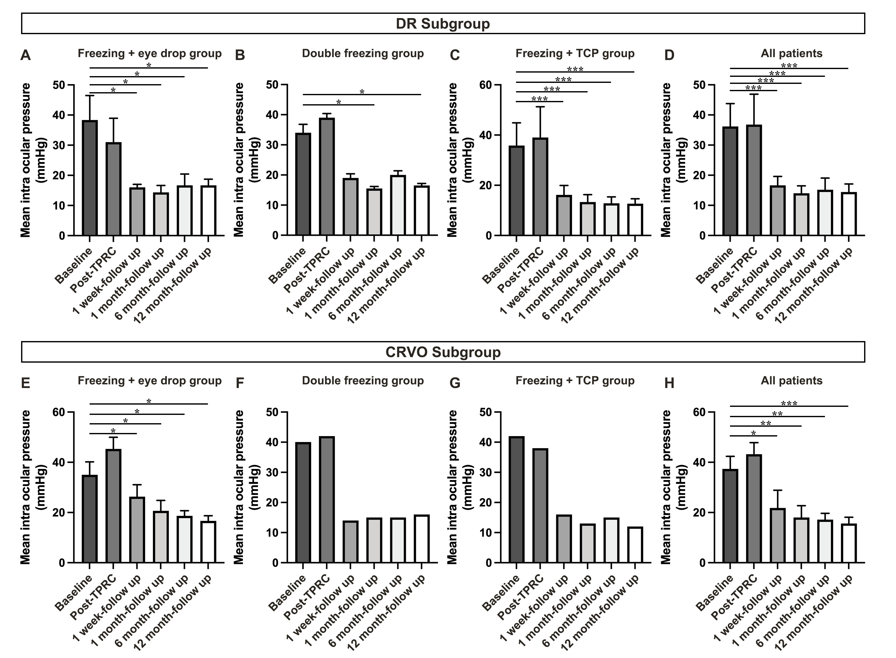
The mean IOP at baseline was 36.56 ± 6.73 mmHg (95% CI: 33.16–39.97). Post-TPRC, IOP increased in most patients, with an average IOP of 38.81 ± 9.11 mmHg (95% CI: 34.20–43.42) on day 1, but no significant difference was observed compared to baseline (P > 0.05) (Fig. 1B). IOP gradually decreased over time in all patients. However, no patient’s IOP reached normal levels, and additional IOP-lowering treatments were required. Based on IOP levels and patient preferences, 6 patients received IOP-lowering eye drops, 3 received cyclocryotherapy, and 6 underwent TCP. At the 1-week follow-up, the IOP was 21.17 ± 6.43 mmHg (95% CI: 17.91–24.42) in the eye drop group, 17.33 ± 3.06 mmHg (95% CI: 15.79–18.88) in the double-freezing group, and 15.79 ± 3.44 mmHg (95% CI: 14.05–17.53) in the TPRC + TCP group (Fig. 1C-E). Significant IOP reductions were observed in all groups (P < 0.05, P < 0.05, P < 0.001). No significant changes in IOP were observed at 1 month, 6 months, or 12 months in any treatment group (P > 0.05). The IOP changes within different original disease subgroup (DR and CRVO) showed similar results (Supplementary Fig. 1A-H).
Long-term IOP changes
For the TPRC + eyedrop group, mean IOP at 1 month, 6 months, and 12 months was 17.50 ± 4.76 mmHg (95% CI: 15.09–19.91), 17.17 ± 3.25 mmHg (95% CI: 15.52–18.81), and 16.67 ± 1.86 mmHg (95% CI: 15.72–17.61), respectively. In the double-freezing group, IOP was 15.33 ± 0.58 mmHg (95% CI: 15.04–15.62) at 1 month, increasing to 18.33 ± 3.06 mmHg (95% CI: 16.79–19.88) at 6 months, and remaining stable at 16.33 ± 0.58 mmHg (95% CI: 15.04–15.62) at 12 months. For the TPRC + TCP group, mean IOP was 13.00 ± 2.69 mmHg (95% CI: 11.64–14.37) at 1 month, 12.92 ± 2.48 mmHg (95% CI: 11.67–14.18) at 6 months, and 12.44 ± 1.81 mmHg (95% CI: 11.53–13.36) at 12 months. No significant difference in IOP was observed among these time points for each group (P > 0.05).
BCVA outcomes
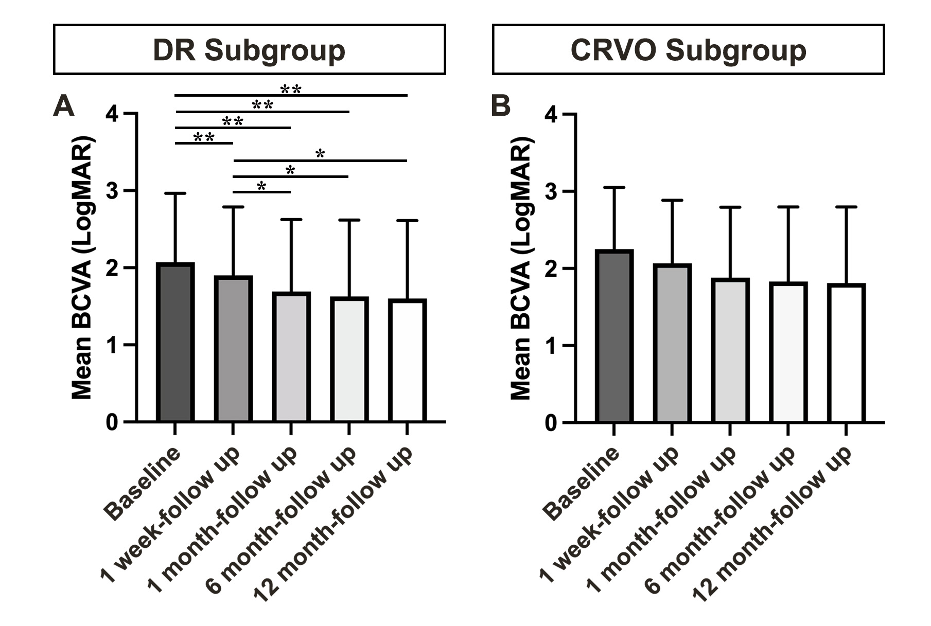
At the 1-week follow-up, BCVA was as follows: 1 patient (6.25%) had NLP, 5 (31.25%) had LP, 2 (12.5%) had HM, 4 (25%) had CF, and 4 (25%) had BCVA better than CF (Fig. 2A). Compared to baseline, 7 patients’ BCVA remained unchanged, while 9 patients showed improvement at the 1-week follow-up. At the 12-month follow-up, 6 patients had unchanged VA, and 10 patients showed improvement (Fig. 2B). No patient experienced a reduction in BCVA. The mean BCVA (in LogMAR) at baseline was 2.25 ± 0.8 (95% CI: 1.84–2.66), improving to 2.07 ± 0.82 (95% CI: 1.66–2.48) at 1 week (P = 0.002). At 1 month, 6 months, and 12 months, the mean BCVA was 1.88 ± 0.91 (95% CI: 1.42–2.43), 1.83 ± 0.97 (95% CI: 1.34–2.32), and 1.81 ± 0.50 (95% CI: 1.31–2.31), respectively, indicating continued improvement (Fig. 2C). The subgroup analysis presented similar result in DR subgroup (Supplementary Fig. 2A). However, due to the small sample size, no significant difference was found in CRVO subgroup (Supplementary Fig. 2B).
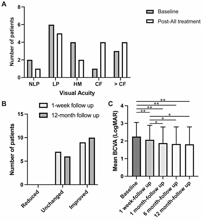
BCVA-Related Results. (A) Distribution of patients with varying BCVA pre- and post-treatment. (B) Number of patients with BCVA changes at 1-week and 12-month follow-up. (C) Mean LogMAR BCVA values at different time points. *P < 0.05; **P < 0.01; ***P < 0.001. Abbreviations: BCVA, best corrected visual acuity; NLP, no light perception; LP, light perception; HM, hand movement; CF, counting fingers
NVI and complications
Iris neovascularization regressed in all patients following TPRC, with complete resolution observed in 2 patients at the 1-week follow-up. No serious complications, such as vision loss, suprachoroidal hemorrhage, retinal detachment, sustained ocular pain, or hypotony, were reported during the follow-up period. However, short-term complications included conjunctival congestion and chemosis (100%), corneal edema (12.5%), corneal epithelial injury (25%), hyphema (6.25%), and anterior chamber exudation (12.5%) (Table 2). All of these complications resolved during subsequent follow-up. Long-term complications included reincrease of iris neovascularization (18.75%), new vitreous hemorrhage (12.5%), and new cataract formation (12.5%) (Table 2).
Discussion
Despite the availability of numerous therapeutic approaches, the management of NVG remains a significant clinical challenge [14]. NVG is a secondary glaucoma driven by underlying pathological conditions that are often difficult to eliminate. Effective management requires minimizing neovascularization and achieving optimal IOP control. This study retrospectively analyzed 16 open-angle NVG cases to evaluate the safety and efficacy of TPRC combined with CAT in controlling IOP and improving VA, offering a promising treatment strategy for NVG.
Currently, numerous treatments have been introduced for managing NVG. Due to the challenges in controlling IOP with conventional medications, invasive procedures are often necessary for NVG patients. Intravitreal injection of anti-VEGF drugs is one of the simple and effective ways. Recent studies indicate that intravitreal injection of bevacizumab can significantly alleviate NVI and maintain normal IOP in 60% of patients for three months [15]. However, the drawbacks of this treatment are evident, as patients must contend with high drug costs and the need for regular visits to medical facilities for injections. Additionally, the risk of intraocular inflammation associated with multiple injections must be taken into account. Furthermore, aqueous drainage surgery is another option for NVG treatment, which includes trabeculectomy and glaucoma drainage device (GDD) implantation surgery. A recent randomized controlled clinical trial related to NVG treatment demonstrated no significant difference in IOP, VA, and complications between patients undergoing GDD implantation and those receiving traditional trabeculectomy. At a one-year follow-up, the success rate of both surgeries was around 60% [16]. At the one-year follow-up, the success rates for both surgical methods were approximately 60%. However, several complications warrant consideration, including early postoperative low IOP, intraocular bleeding, long-term scar formation of the filtering bleb, and exposure of the GDD. This necessitates close postoperative and long-term follow-up for patients. The combination therapy based on TPRC introduced in this study requires simple equipment and low technical skills for operators, yet it offers a high postoperative IOP control rate, thereby mitigating some of the shortcomings associated with the aforementioned treatments. A previous meta-analysis indicated no significant difference in efficacy between TCP and GDD implantation surgery [11], suggesting that better leveraging the advantages of the combined therapy of cycloablation and TPRC treatments may be beneficial.
Retinal ischemia due to vascular occlusion underpins NVG pathogenesis. While reperfusion of occluded retinal vessels remains unattainable, reducing the reactivity of ischemic retinal cells is critical. PRP, as the standard treatment, addresses retinal ischemia and improves NVG prognosis [10]. However, PRP is limited to patients with clear dioptric media and cannot effectively target the extreme periphery retina. Although widely utilized [5], PRP alone often fails to adequately control IOP, necessitating supplementary therapies.
TPRC complements PRP by addressing peripheral ischemic retinal areas inaccessible to laser. Early studies validate the efficacy of retinal cryotherapy in NVG management. In 1980, May et al. reported that TPRC reduced IOP to manageable levels within three days, with NVI regressing within six weeks [17]. Vernon et al. (1988) observed regression of NVI, stable VA, and normalized IOP in 8 of 9 eyes treated with TPRC [18]. Subsequent studies reinforced these findings, demonstrating improved VA, reduced IOP, and significant NVI regression in patients treated with TPRC, either alone or combined with limited cyclocryotherapy [19, 20]. These studies collectively highlight the potential of cryotherapy in mitigating NVG progression through NVI suppression and VEGF reduction.
While TPRC alone reduces NVI, achieving sustained IOP control often requires adjunctive therapies. Combination approaches, such as TPRC with trabeculectomy or TCP, have shown improved outcomes. For instance, Vigo et al. (1988) reported IOP control in 75% of NVG patients treated with TPRC and trabeculectomy [21]. More recent studies suggest that integrating TPRC with intravitreal bevacizumab or TCP further enhances IOP control and VA stability [22, 23].
In this study, TPRC combined with CAT demonstrated effectiveness in reducing IOP and improving VA with several advantages: the procedure is relatively simple, cost-effective, and avoids reliance on consumables such as glaucoma valves or the risk of filtration channel closure associated with trabeculectomy. This approach is particularly suited for NVG patients with extremely elevated IOP, where immediate intervention to lower IOP is critical to prevent optic nerve damage, the primary cause of blindness in these patients [24]. In this study, 10 out of 16 patients (62.5%) received additional CAT treatment. In contrast, May et al. reported a rate of 83.3% in their previous study [17], while Brodell et al. found a rate of 48.1% [19]. Although our findings indicate a relatively median rate, it is evident that further CAT treatment remains essential after TPRC for at least half of NVG patients. Additionally, previous research has confirmed that combining TPRC with cyclocryotherapy yields better outcomes than cyclocryotherapy alone [25]. These results suggest that NVG often necessitates a multifaceted approach to achieve adequate IOP control and mitigate neovascularization.
Cycloablation techniques like TCP, while effective for IOP reduction, are associated with complications such as hyphema, hypotony, and vision loss [26,27,28]. To minimize these risks, a staged approach was employed in this study: TPRC was initially used to reduce NVI, followed by graduated cycloablation therapy. This protocol allowed for gradual IOP reduction and a better safety profile, with no hypotony or significant hyphema observed at 12 months. In this study, VA improvement was observed in 56.25% (9/16) of patients at the early stages following treatment, and in 62.5% (10/16) of patients at the later stages. Among patients with early VA improvement, 7 patients experienced significant relief from corneal edema, while the remaining 2 benefited from the alleviation of optic nerve ischemia due to a decrease in IOP. The long-term improvement in VA may stem from the restoration of blood supply to the optic nerve and retina after sustained IOP reduction. Although the endpoint follow-up time was established at 12 months, a recent telephone follow-up with 12 patients (4 patients were lost to follow-up) indicated that long-term maintenance of normal IOP was achieved in all patients. The patient who maintained normal IOP for the longest duration has surpassed 4 years, while the shortest duration has exceeded 2 years. None of these 12 patients experienced serious complications, demonstrating the safety and effectiveness of the TPRC-based combination therapy. The dynamic balance between the existing functional trabecular meshwork and aqueous humor secretion is crucial for the long-term stability of IOP in this therapy. For all included patients, we conducted thorough patient education and encouraged them to actively manage their blood pressure and sugar levels to minimize the risk of further development of NVG. This is particularly important for patients with NVG resulting from DR. Therefore, effective doctor-patient cooperation is also a potential key factor in the success of this therapy.
Careful titration of cycloablation parameters and observation periods between treatments were key to achieving these outcomes. Short-term IOP-lowering eye drops provided transitional support during these intervals, ensuring adequate IOP control while awaiting the full effects of treatment.
This study presents several limitations. The small sample size and narrow etiology, limited to PDR and CRVO, may introduce selection bias. Typically, a retrospective study on glaucoma treatment should involve more than 50 participants [29]. While early studies on TPRC treatment from the last century included only about 10 patients [17, 18], a small sample size can still lead to significant bias in research outcomes. Additionally, the relatively short follow-up period limits the assessment of long-term IOP control and VA stability. As a retrospective analysis, this study lacks the rigor of a standardized prospective trial, precluding direct comparisons with other treatment modalities. Despite these limitations, the findings suggest that TPRC combined with CAT offers a viable option for primary care settings or resource-limited hospitals. This approach is particularly suitable for NVG patients with extreme IOP elevations and limited economic means. Future prospective, multicenter studies with larger sample sizes and extended follow-up periods are warranted to further validate this treatment strategy and explore its comparative efficacy against other NVG management options.
Conclusion
This study conducted a retrospective observational case series to describe and evaluate the efficacy of TPRC combined with other IOP-lowering treatments in patients with open-angle NVG. Results confirmed the IOP-controlling and VA-preserving effect of TPRC-based therapy for at least 1 year. These TPRC-based IOP-lowering combination therapies appear to be an effective treatment option for open-angle NVG. They are particularly suitable for primary healthcare settings, hospitals without advanced glaucoma surgery capabilities, or NVG patients with limited economic resources and extremely elevated IOP.
Data availability
The raw datasets are available on the website as supplementary files. More datasets analysed during the current study are available from the corresponding author on reasonable request.
Abbreviations
- BCVA:
-
Best corrected visual acuity
- CAT:
-
Cycloablation treatment
- CI:
-
Confidence interval
- DR:
-
Diabetic retinopathy
- GDD:
-
Glaucoma drainage device
- iCRVO:
-
Ischemic central retinal vein occlusion
- IOP:
-
Intraocular pressure
- NVG:
-
Neovascular glaucoma
- NVI:
-
Iris neovascularization
- OIS:
-
Ocular ischemic syndrome
- PDR:
-
Proliferative diabetic retinopathy
- PRP:
-
Pan-retinal photocoagulation
- SD:
-
Standard deviation
- VA:
-
Visual acuity
- VEGF:
-
Vascular endothelial growth factor
References
Narayanaswamy A, et al. The prevalence and types of glaucoma in an urban Indian population: the Singapore Indian eye study. Invest Ophthalmol Vis Sci. 2013;54(7):4621–7.
Wong TY, et al. Prevalence and causes of low vision and blindness in an urban Malay population: the Singapore Malay eye study. Arch Ophthalmol. 2008;126(8):1091–9.
Senthil S, et al. Neovascular glaucoma - A review. Indian J Ophthalmol. 2021;69(3):525–34.
Ohrt V. The frequency of rubeosis Iridis in diabetic patients. Acta Ophthalmol (Copenh). 1971;49(2):301–7.
Kim YH, Sung MS, Park SW. Clinical features of ocular ischemic syndrome and risk factors for neovascular Glaucoma. Korean J Ophthalmol. 2017;31(4):343–50.
Yang H, Yu X, Sun X. Neovascular glaucoma: handling in the future. Taiwan J Ophthalmol. 2018;8(2):60–6.
Mocanu C, et al. [Neovascular glaucoma–retrospective study]. Oftalmologia. 2005;49(4):58–65.
Chalam KV, Brar VS, Murthy RK. Human ciliary epithelium as a source of synthesis and secretion of vascular endothelial growth factor in neovascular glaucoma. JAMA Ophthalmol. 2014;132(11):1350–4.
Urbonaviciute D, Buteikiene D, Januleviciene I. A review of neovascular glaucoma: etiology, pathogenesis, diagnosis, and treatment. Med (Kaunas). 2022;58(12).
Havens SJ, Gulati V. Neovascular Glaucoma Dev Ophthalmol. 2016;55:196–204.
Shchomak Z, et al. Surgical treatment of neovascular glaucoma: a systematic review and meta-analysis. Graefes Arch Clin Exp Ophthalmol. 2019;257(6):1079–89.
Rani PK, et al. Outcomes of neovascular glaucoma in eyes presenting with moderate to good visual potential. Int Ophthalmol. 2021;41(7):2359–68.
Day AC, et al. The Royal college of ophthalmologists’ National ophthalmology database study of cataract surgery: report 1, visual outcomes and complications. Eye (Lond). 2015;29(4):552–60.
Dong Z, et al. Effectiveness of multiple therapeutic strategies in neovascular glaucoma patients: A PRISMA-compliant network meta-analysis. Med (Baltim). 2018;97(14):e9897.
Palfi Salavat MC, et al. The role of Anti-VEGF agents in treatment of neovascular glaucoma. Rom J Ophthalmol. 2022;66(3):209–13.
Tokumo K, et al. Treatment outcomes in the neovascular glaucoma tube versus trabeculectomy study. Graefes Arch Clin Exp Ophthalmol. 2021;259(10):3067–76.
May DR, et al. Treatment of neovascular glaucoma with transscleral panretinal cryotherapy. Ophthalmology. 1980;87(11):1106–11.
Vernon SA, Cheng H. Panretinal cryotherapy in neovascular disease. Br J Ophthalmol. 1988;72(6):401–5.
Brodell LP, et al. Neovascular glaucoma: a retrospective analysis of treatment with peripheral panretinal cryotherapy. Ophthalmic Surg. 1987;18(3):200–6.
Chen T, et al. [The change of the level of the vascular endothelial growth factor in aqueous humor of patients with neovascular glaucoma before and after anterior retinal cryotherapy]. Zhonghua Yan Ke Za Zhi. 2007;43(7):622–5.
Fernandez-Vigo J, et al. Treatment of diabetic neovascular glaucoma by panretinal ablation and trabeculectomy. Acta Ophthalmol (Copenh). 1988;66(6):612–6.
Kuang TM, et al. Clinical experience in the management of neovascular glaucoma. J Chin Med Assoc. 2004;67(3):131–5.
Tatsumi T, et al. Panretinal photocoagulation with simultaneous cryoretinopexy or intravitreal bevacizumab for neovascular glaucoma. Graefes Arch Clin Exp Ophthalmol. 2013;251(5):1355–60.
Hayreh SS. Neovascular glaucoma. Prog Retin Eye Res. 2007;26(5):470–85.
Rehak J, Vymazal M. [Cryotherapy in treatment of neovascular glaucoma with closed chamber angle]. Klin Monbl Augenheilkd. 1994;204(1):20–3.
Freigassner P, Eckhardt M. Transscleral cyclophotocoagulation versus cyclocryotherapy in treatment of neovascular glaucoma: a retrospective analysis. Acta Ophthalmol Scand. 2003;81(6):674–5.
Leszczynski R, et al. Contact transscleral cyclophotocoagulation in the treatment of neovascular glaucoma: a five-year follow-up. Med Sci Monit. 2009;15(3):BR84–7.
Scott TM, et al. Long-term limitations and complications of trans-scleral diode laser cycloablation for refractory glaucoma. Clin Exp Ophthalmol. 2023;51(2):131–6.
Bernal-Morales C, et al. Simultaneous Pars plana vitrectomy, panretinal photocoagulation, cryotherapy, and Ahmed valve implantation for neovascular glaucoma. Int J Ophthalmol. 2021;14(9):1396–401.
Acknowledgements
NA.
Funding
NA.
Author information
Authors and Affiliations
Contributions
LXG and JFH performed the preparation, analyzed the data and wrote the manuscript. LQC, LYR, BC, and XKZ collect the data. LXG and WYS prepared figures and Tables. WYS was responsible for revising the paper. FXW was responsible for the supervision of the paper, revision of the drafts, and the final reversion of the paper. All authors read and approved the submitted manuscript.
Corresponding authors
Ethics declarations
Ethics approval and consent to participate
All data of this study was retracted from the medical records of patients. The written informed consent for the use of clinical data in future retrospective studies from each participant was obtained. The Institutional Review Board of the PLA General Hospital reviewed and approved the study protocol. This study was performed in accordance with the ethical standards stated in the 2013 Declaration of Helsinki.
Consent for publication
Not applicable.
Competing interests
The authors declare no competing interests.
Additional information
Publisher’s note
Springer Nature remains neutral with regard to jurisdictional claims in published maps and institutional affiliations.
Electronic supplementary material
Below is the link to the electronic supplementary material.


Rights and permissions
Open Access This article is licensed under a Creative Commons Attribution-NonCommercial-NoDerivatives 4.0 International License, which permits any non-commercial use, sharing, distribution and reproduction in any medium or format, as long as you give appropriate credit to the original author(s) and the source, provide a link to the Creative Commons licence, and indicate if you modified the licensed material. You do not have permission under this licence to share adapted material derived from this article or parts of it. The images or other third party material in this article are included in the article’s Creative Commons licence, unless indicated otherwise in a credit line to the material. If material is not included in the article’s Creative Commons licence and your intended use is not permitted by statutory regulation or exceeds the permitted use, you will need to obtain permission directly from the copyright holder. To view a copy of this licence, visit http://creativecommons.org/licenses/by-nc-nd/4.0/.
About this article

Cite this article
Gao, L., Huang, J., Cao, L. et al. Effect of combined therapy based on transscleral peripheral retinal cryotherapy in the treatment of open-angle neovascular glaucoma: a retrospective study. BMC Ophthalmol 25, 375 (2025). https://doi.org/10.1186/s12886-025-04185-7
Received:
Accepted:
Published:
Version of record:
DOI: https://doi.org/10.1186/s12886-025-04185-7