

Abstract
Background
Mixed parasitic infections are prevalent in dogs, often leading to significant clinical issues and posing zoonotic risks to humans. Dogs can simultaneously harbor ectoparasites such as fleas and ticks and endoparasites such as gastrointestinal nematodes. These parasites can induce dermatological and gastrointestinal disorders and serve as vectors or reservoirs for zoonotic infections. This case report describes a rare triple parasitic co-infection involving Ctenocephalides canis, Rhipicephalus sanguineus, and Toxocara canis in a domestic dog in Bangladesh and highlights the diagnostic approach and effective treatment strategy.
Methods
A 2-year-old male local breed dog presented to the veterinary clinic at Bangladesh Agricultural University, Mymensingh, Bangladesh, exhibiting severe pruritus, reduced body condition score, and enteric signs. Parasites were collected and identified through gross examination and detailed morphological analysis under a stereomicroscope (Olympus SZ61, Olympus Corporation, Tokyo, Japan). Key diagnostic features included cervical alae in T. canis, genal and pronotal combs in C. canis, and a hexagonal basis capitulum in R. sanguineus. Treatment consisted of an oral combination of sarolaner (1200 µg/kg), moxidectin (24 µg/kg), and pyrantel pamoate (5000 µg/kg), accompanied by supportive therapy and environmental control measures.
Main results
Morphological examination identified 35 adult Ctenocephalides canis, 12 adult Rhipicephalus sanguineus and 4 adult Toxocara canis, thereby indicating a concurrent triple infection. Following treatment, clinical improvement was observed within 48 h, characterized by a marked reduction in pruritus and improved appetite. A follow-up examination at 14 days revealed no visible ectoparasites and a negative fecal flotation test for T. canis eggs. Complete clinical recovery was recorded during the 30-day follow-up period.
Conclusions
This case underscores the clinical and zoonotic significance of mixed parasitic infections in dogs. Routine parasitic screening and preventive measures are crucial within a One Health framework to minimize zoonotic transmission risks.
Introduction
Mixed parasitic infections are frequently observed in dogs, carrying significant clinical and zoonotic implications (Tran Nguyen-Ho- et al. 2023, Macedo et al. 2022). Dogs can harbor a variety of ectoparasites, such as fleas (Ctenocephalides canis, Pulex irritans), ticks (Rhipicephalus sanguineus, Amblyomma americanum), and mites (Demodex canis, Sarcoptes scabiei), as well as endoparasites like roundworms (Toxocara canis, Ancylostoma caninum) and tapeworms (Dipylidium caninum) (Ortega-Pacheco and Jimenez-Coello 2025). These parasites not only inflict direct harm upon the host, leading to conditions such as pruritus, reduced body condition score, and enteric signs, but also serve as vectors for various pathogens, thereby posing a public health risk (Alba-Hurtado et al. 2022, Despommier and Toxocariasis 2003). For instance, Rhipicephalus sanguineus is known to transmit several zoonotic microorganisms (Do et al. 2024, Ghodrati et al. 2024), and Toxocara canis is a well-recognized cause of toxocariasis in humans (Yang et al. 2025, Reginaldo et al. 2025). The impact of co-infection interactions on parasite transmission dynamics can vary with spatial scale (Keegan et al. 2024).
Previous studies have reported considerable prevalence of canine ectoparasites and endoparasites in South Asia. In Bangladesh, studies have shown ectoparasite infestation rates ranging from 59.57% to 64% in dogs, with Ctenocephalides canis and Rhipicephalus sanguineus being predominant species (Zineldar et al. 2023, Sadia Authoy et al. 2023). Concurrently, gastrointestinal parasitic infections are highly prevalent, with recent studies in Dhaka reporting an overall prevalence of 63.33% in dogs, where Toxocara canis is frequently identified (Bayazid et al. 2025). Although mixed parasitic infections in dogs have been reported globally, published reports describing concurrent infection with Ctenocephalides canis, Rhipicephalus sanguineus, and Toxocara canis in a single host remain scarce. To the best of our knowledge, this is one of the few documented reports of such a specific triple co-infection from Bangladesh, highlighting the potential for simultaneous ecto- and endoparasitic infestations in companion animals within the region. This case report details this triple parasitic co-infection in a domestic dog, emphasizing the diagnostic approach, treatment, and broader implications for animal and human health.
Case history
A 2-year-old male local breed dog presented with severe pruritus, reduced body condition score, and enteric signs. The owner reported a history of persistent pruritus and visible ectoparasites. Further details regarding the dog’s environment, vaccination status, and previous deworming history were collected to establish a comprehensive case profile.
Diagnosis
Fleas and ticks were manually removed from the dog’s body using fine-tipped forceps. Fleas were collected mainly from the dorsal trunk and inguinal regions, whereas ticks were collected from the ears and neck. Adult Toxocara canis worms were recovered from freshly voided feces after spontaneous passage. Each parasite specimen was preserved separately in sterile containers containing 70% ethanol and transported to the laboratory. Before microscopic examination, specimens were rinsed with normal saline, examined grossly, and subsequently evaluated under a stereomicroscope (Olympus SZ61, Olympus Corporation, Tokyo, Japan) for detailed morphological identification.
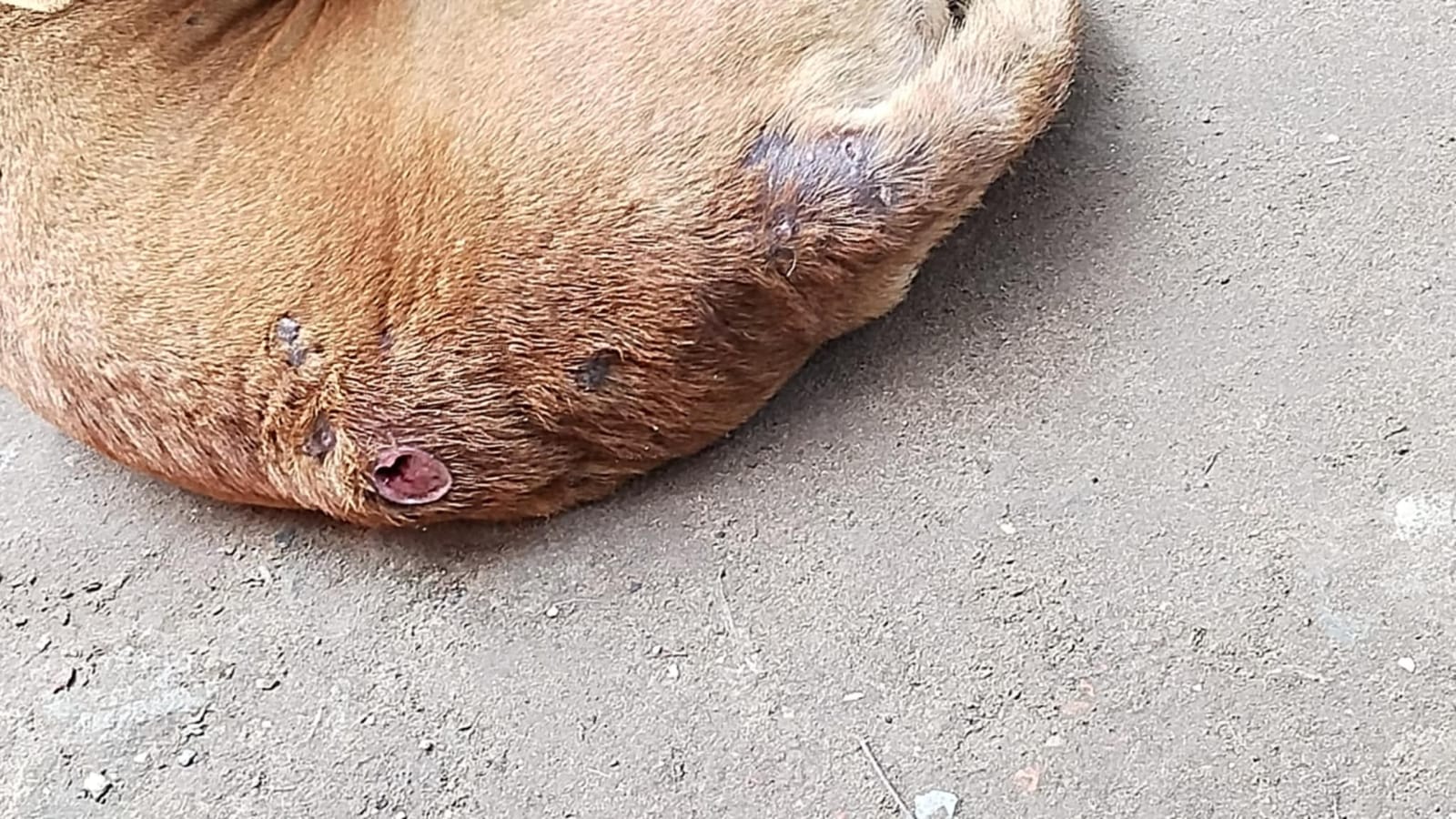
Gross examination revealed multiple attached ectoparasites and localized dermatological lesions in the ventral abdominal region (Supplementary Figure S1). Diagnosis was established through gross and microscopic examinations of parasites collected from the patient. The identification of each parasite species was performed using the standard morphological taxonomic keys described in Helminths, Arthropods and Protozoa of Domesticated Animals, Georgis’ Parasitology for Veterinarians and Ticks of Domestic Animals in Africa: A Guide to Identification of Species (Walker Alan 2003, Bowman and Georgi 2021, ’Soulsby and Helminths 1982). Diagnostic identification was based on established morphological characteristics observed under a stereomicroscope (Olympus SZ61, Olympus Corporation, Tokyo, Japan) and independently verified by a veterinary parasitologist from the Department of Parasitology, Bangladesh Agricultural University (Table 1).
These morphological characteristics (Figs. 1 and 2), particularly the hexagonal basis capituli of R. sanguineus, the specific comb structure of C. canis, and the cervical alae of T. canis, provided definitive evidence for the triple parasitic co-infection and were consistent with previously published taxonomic descriptions (Walker Alan 2003, Bowman and Georgi 2021, ’Soulsby and Helminths 1982). Approximately 35 adult Ctenocephalides canis, 12 adult Rhipicephalus sanguineus and 4 adult Toxocara canis specimens were recovered from the affected dog.

Gross morphological features of Toxocara canis (A), Ctenocephalides canis (B), and Rhipicephalus sanguineus (C) specimens recovered from a domestic dog

This figure provides detailed microscopic views of the parasitic species, crucial for definitive identification. Panel (A) shows the anterior end of Toxocara canis. Panel (B) displays microscopic features of Ctenocephalides canis. Panel (C) illustrates the anterior and Panel (D) the posterior parts of Rhipicephalus sanguineus
Treatment and follow-up of the case and control measures
The patient received a comprehensive antiparasitic regimen designed to simultaneously address both ecto and endoparasitic burdens. Following standard veterinary protocols (Simparica ), the dog was administered a combination product containing sarolaner (1200 µg/kg), moxidectin (24 µg/kg), and pyrantel pamoate (5000 µg/kg) in a single oral dose.
Treatment guidelines and supportive therapy
Antiparasitic administration
The oral tablet was administered with food to ensure optimal absorption. Sarolaner was selected because of its proven efficacy against fleas and ticks, while moxidectin and pyrantel pamoate provide broad-spectrum activity against gastrointestinal nematodes including T. canis (Simparica ). The combination allows simultaneous treatment of ectoparasitic and endoparasitic infections with a single administration. This provides immediate and persistent efficacy against fleas and ticks for one month, concurrently treating the T. canis infection.
Supportive care
To manage severe pruritus and skin irritation, a topical soothing shampoo and a short course of antihistamines were administered. Enteric signs were managed with a highly digestible diet and probiotic supplementation for seven days.
Environmental control
The owner was instructed to thoroughly clean the dog’s living environment and apply a permethrin-based environmental insecticide to eliminate flea larvae, pupae, adult fleas, and off-host tick stages. Environmental treatment is recommended because most stages of the flea life cycle occur away from the host, and environmental decontamination significantly reduces reinfestation and improves long-term parasite control (ESCCAP, Dryden 2009).
Follow-up and recovery
Clinical improvement was observed within 48 h, marked by a significant reduction in pruritus and improved appetite. A follow-up examination at 14 days revealed no visible ectoparasites and a negative fecal flotation test for T. canis eggs. The animal showed complete recovery by the end of the 30-day follow-up period.
Discussion
While mixed infections are frequently suspected in clinical practice, this case offers detailed morphological documentation of a specific triple co-infection, underscoring the necessity for broad-spectrum parasiticides in regions where environmental conditions and stray animal populations favor high parasite diversity. The co-occurrence of Ctenocephalides canis, Rhipicephalus sanguineus, and Toxocara canis underscores the need for comprehensive diagnostic approaches that consider both ecto- and endoparasites. The zoonotic potential of these parasites, particularly R. sanguineus as a vector for various pathogens (Do et al. 2024, Ghodrati et al. 2024, Özübek 2024, Gharbi et al. 2024) and T. canis as a cause of human toxocariasis (Yang et al. 2025, Reginaldo et al. 2025), emphasizes the public health significance of such cases. It is important to note that human exposure risk was not directly assessed through household surveys or diagnostic testing in this study. The zoonotic implications discussed in this report are based on the established zoonotic potential of T. canis and pathogen transmission associated with R. sanguineus as reported in the literature (Alba-Hurtado et al. 2022, Do et al. 2024, Keegan et al. 2024). Furthermore, although no recurrence was observed during the 30-day follow-up period, longer monitoring may be necessary to exclude the possibility of reinfection or delayed relapse, particularly in environments with persistent parasite contamination.
Molecular confirmation of parasite identity through PCR amplification and DNA sequencing was not performed because molecular diagnostic facilities and research funding were unavailable during this case investigation. Nevertheless, morphological identification was considered reliable because all recovered specimens exhibited the characteristic diagnostic features described in internationally accepted taxonomic keys (Bowman and Georgi 2021). Future studies should incorporate molecular techniques to further validate species identification and investigate parasite genetic diversity.
Effective control strategies must therefore encompass both animal treatment and environmental management, aligning with the ‘One Health’ perspective (Giannelli et al. 2024). Studies have shown the epidemiological characteristics of ectoparasite infestation in domestic dogs (Tran Nguyen-Ho- et al. 2023, Zineldar et al. 2023, Sadia Authoy et al. 2023) and the prevalence of potentially zoonotic endoparasites (Reginaldo et al. 2025, Bayazid et al. 2025). These findings are consistent with previous reports from Bangladesh, where Ctenocephalides canis and Rhipicephalus sanguineus have been identified as the predominant ectoparasites of domestic dogs (Sadia Authoy et al. 2023), while Toxocara canis remains one of the most frequently detected zoonotic gastrointestinal nematodes (Bayazid et al. 2025). The present report extends these observations by documenting the concurrent occurrence of all three parasite species in a single dog. The impact of co-infection interactions on between-host parasite transmission dynamics also represents a critical area of research (Keegan et al. 2024, Hananeh et al. 2022, Milićević et al. 2025). Future investigations should employ polymerase chain reaction (PCR) assays followed by sequencing of mitochondrial cytochrome-c oxidase subunit I (cox1), 16 S rRNA and Internal Transcribed Spacer (ITS) gene regions to confirm parasite identity, evaluate genetic diversity, detect cryptic species and identify associated vector-borne pathogens that may not be distinguishable through morphology alone (Nyema et al. 2024, Zazueta-Islas et al. 2024).
From a One Health perspective, routine deworming of dogs, year-round flea and tick control, responsible pet ownership, proper disposal of animal feces, public education regarding zoonotic parasites and coordinated surveillance involving veterinarians and public health authorities are recommended to reduce transmission risks at the community level.
Conclusions
This case report documents a significant instance of triple parasitic co-infection involving Ctenocephalides canis, Rhipicephalus sanguineus, and Toxocara canis in a domestic dog. The successful management of this case through integrated diagnostic and therapeutic protocols highlights the critical need for vigilance in veterinary practice. Crucially, this case emphasizes the profound zoonotic importance of these parasites. Toxocara canis poses a direct threat to human health through visceral and ocular larva migrans, while Rhipicephalus sanguineus serves as a primary vector for life-threatening zoonotic diseases such as Rocky Mountain spotted fever and ehrlichiosis. Therefore, routine parasitic screening and the implementation of year-round preventive measures are not merely animal health concerns but constitute essential public health interventions. A collaborative ‘One Health’ approach involving pet owners, veterinarians, and public health officials is vital to mitigate the risks of zoonotic transmission in urban and rural environments.
Data availability
The datasets generated and analyzed during the current study are available from the corresponding author on reasonable request.
Change history
01 August 2026
The original publication was amended to update the Acknowledgements.
References
Alba-Hurtado F, Antonio Muñoz-Guzmán M (2022) Toxocariosis: from a one health perspective. In: Parasitic helminths and zoonoses - from basic to applied research. IntechOpen. https://doi.org/10.5772/intechopen.104508
Bayazid A, Al H, Sutradhar MF, Nahar S, Harun A, Bin A, Sultana T et al (2025) Prevalence, risk factors, and zoonotic implications of gastrointestinal parasites in dogs and cats in Dhaka City, Bangladesh. Vet Parasitol Reg Stud Rep 65:101358. https://doi.org/10.1016/j.vprsr.2025.101358
Bowman DD, Georgi JR (2021) Georgis’ parasitology for veterinarians. Elsevier, p 518
CDC DPDx DPDx - Laboratory identification of parasites of public health concern search. https://www.cdc.gov/dpdx/. Accessed 17 Jul 2026
de Macedo LO, Bezerra-Santos MA, Filho CRCU, da Silva Sales KG, de Sousa-Paula LC, da Silva LG et al (2022) Vector-borne pathogens of zoonotic concern in dogs from a Quilombola community in northeastern Brazil. Parasitol Res 121(11):3305–3311. https://doi.org/10.1007/s00436-022-07661-x
Despommier D, Toxocariasis (2003) Clinical Aspects, Epidemiology, Medical Ecology, and Molecular Aspects. Clin Microbiol Rev 16(2):265–272. https://doi.org/10.1128/CMR.16.2.265-272.2003
Do T, Bui LK, Umemiya-Shirafuji R, Inpankaew T, Hasan T, Zafar I et al (2024) The detection of zoonotic microorganisms in Rhipicephalus sanguineus (brown dog ticks) from Vietnam and the frequency of tick infestations in owned dogs. Front Vet Sci 11. https://doi.org/10.3389/fvets.2024.1435441
Dryden MW (2009) Flea and tick control in the 21st century: challenges and opportunities. Vet Dermatol 20(5–6):435–440. https://doi.org/10.1111/j.1365-3164.2009.00838.x
ESCCAP ESCCAP Guideline 03. https://www.esccap.org/uploads/docs/ise18g5s_ESCCAP_Guideline_03_Third_Edition__September_2015.pdf. Accessed 17 Jul 2026
Gharbi Z, Ouni A, Balti G, Bouattour A, Chabchoub A, M’ghirbi Y (2024) First Evidence of Rickettsia conorii Infection in Dogs in Northern Tunisia. Vet Sci 11(9):402. https://doi.org/10.3390/vetsci11090402
Ghodrati S, Lesiczka PM, Zurek L, Szekely F, Modrý D (2024) Rhipicephalus sanguineus from Hungarian dogs: Tick identification and detection of tick-borne pathogens. Vet Parasitol Reg Stud Rep 50:101007. https://doi.org/10.1016/j.vprsr.2024.101007
Giannelli A, Schnyder M, Wright I, Charlier J (2024) Control of companion animal parasites and impact on One Health. One Health 18:100679. https://doi.org/10.1016/j.onehlt.2024.100679
Hananeh WM, Radhi A, Mukbel RM, Ismail ZB (2022) Effects of parasites coinfection with other pathogens on animal host: a literature review. Vet World 2414–2424. https://doi.org/10.14202/vetworld.2022.2414-2424
Keegan SP, Pedersen AB, Fenton A (2024) The impact of within-host coinfection interactions on between-host parasite transmission dynamics varies with spatial scale. Proc R Soc B Biol Sci 291(2021). https://doi.org/10.1098/rspb.2024.0103
Milićević V, Mira F, Kavran M, Editorial (2025) Sentinels of health: advancements in monitoring and surveillance of vector-borne diseases in domestic and wild animals and vectors. Front Vet Sci. https://doi.org/10.3389/fvets.2025.1670316
Nyema J, Nath TC, Bhuiyan MJU, Islam S, Islam KM, Basir S et al (2024) Morpho-molecular investigation of ectoparasitic infestation of companion animals in Sylhet city, Bangladesh. Vet Parasitol Reg Stud Rep 47:100953. https://doi.org/10.1016/j.vprsr.2023.100953
Özübek S (2024) Preliminary Study on the Genetic Diversity of Hepatozoon canis in Dogs and Rhipicephalus sanguineus sensu lato Ticks. Dicle Üniversitesi Veteriner Fakültesi Dergisi 17(2):118–123. https://doi.org/10.47027/duvetfd.1504235
Ortega-Pacheco A, Jimenez-Coello M (2025) Vector-Borne and Zoonotic Diseases in Dogs and Cats. Animals 15(11):1520. https://doi.org/10.3390/ani15111520
Reginaldo GMdos, Widmer S, Inácio G, Gomes SV, Bertequini Nagata JF, Moreno W (2025) Prevalence of Potentially Zoonotic Endoparasites in Domestic Dog Puppies. Vet Sci 12(4):332. https://doi.org/10.3390/vetsci12040332
Sadia Authoy A, Chanda A, Datta A, Al Faruk MS, Kamal T (2023) Prevalence of ectoparasites in pet dog and cat in Dhaka, Bangladesh. Res J Vet Pract 11(4). https://doi.org/10.17582/journal.rjvp/2023/11.4.42.48
Simparica Trio (sarolaner, moxidectin, and pyrantel chewable tablets): Prescribing information (2020) https://dailymed.nlm.nih.gov/dailymed/drugInfo.cfm?setid=0f83bcf4-7e89-479f-a980-4cb40456ee78. Accessed 2 Mar 2026
’Soulsby E, Helminths (1982) Arthropods and protozoa of domesticated animals. 7th ed. London: Baillière Tindall. https://www.journeywithasr.com/2021/01/helminths-arthropods-and-protozoa-of-domesticated-animals-by-ejl-soulsby.html. Accessed 17 Jul 2026
Tran Nguyen-Ho-, Bao, Thi La MA, Nguyen BP, Nguyen CT, Nguyen TM, Lu TA et al (2023) Epidemiological characteristics of the ectoparasite infestation in domestic dogs in the Mekong Delta of Vietnam. Vet Integr Sci 22(3). https://doi.org/10.12982/VIS.2024.078
Walker Alan (2003) Ticks of domestic animals in Africa: a guide to identification of species. Biosci Rep 221 p https://www.researchgate.net/publication/291049181_Ticks_of_Domestic_Animals_in_Africa_a_guide_to_identification_of_species
Yang Y, Zhong D, Wu H, Xiong Z, Yang C, Yang L et al (2025) Prevalence of Toxocara infection and associated risk factors: a cross-sectional study in Zhejiang, China. Infect Dis Poverty 14(1):43. https://doi.org/10.1186/s40249-025-01312-w
Zazueta-Islas HM, Salceda-Sánchez B, Huerta-Jiménez H, Miranda-Caballero CI, Solis-Cortés M, de la Cruz-Pacheco Y et al (2024) Molecular Detection of Kinetoplastid Species in Ticks and Fleas Associated with Dogs and Humans in Mexico. Pathogens 13(12):1072. https://doi.org/10.3390/pathogens13121072
Zineldar H, Zeid N, Eisa M, Bennour E, Neshwey W (2023) Prevalence, clinical presentation, and therapeutic outcome of ectoparasitic infestations in dogs in Egypt. Open Vet J 13(12):1631. https://doi.org/10.5455/OVJ.2023.v13.i12.13
Acknowledgements
The author sincerely thanks the owner of the dog for consenting to the clinical examination and treatment of the animal. The author also gratefully acknowledges Dr. A. R. M. Beni Amin for collecting the samples, treating the dog and documenting the case through photographs. The author further acknowledges the Department of Parasitology and the Veterinary Teaching Hospital, Bangladesh Agricultural University, Mymensingh, for providing laboratory and clinical support during the diagnosis and management of this case.
Funding
This research did not receive any specific grant from funding agencies in the public, commercial or not-for-profit sectors.
Author information
Authors and Affiliations
Contributions
Tanvir Ahmmed Joy: Conceived and designed the study, performed data analysis, methodological development, data interpretation, critical revision and edited the manuscript.
Corresponding author
Ethics declarations
Ethics approval and consent to participate
Ethical approval was not required for this case report according to institutional guidelines, as all diagnostic and therapeutic procedures were performed as part of routine veterinary clinical care without any experimental intervention. The study complied with accepted standards of veterinary clinical practice. Written informed consent was obtained from the owner of the dog for clinical examination, treatment, collection of parasite specimens for diagnostic purposes, and publication of this case report and accompanying images.
Consent for publication
The author has read and approved the final version of the manuscript and consents to its publication.
Competing interests
The authors declare no competing interests.
Additional information
Publisher’s Note
Springer Nature remains neutral with regard to jurisdictional claims in published maps and institutional affiliations.
Supplementary Information
Below is the link to the electronic supplementary material.
Rights and permissions
Open Access This article is licensed under a Creative Commons Attribution 4.0 International License, which permits use, sharing, adaptation, distribution and reproduction in any medium or format, as long as you give appropriate credit to the original author(s) and the source, provide a link to the Creative Commons licence, and indicate if changes were made. The images or other third party material in this article are included in the article’s Creative Commons licence, unless indicated otherwise in a credit line to the material. If material is not included in the article’s Creative Commons licence and your intended use is not permitted by statutory regulation or exceeds the permitted use, you will need to obtain permission directly from the copyright holder. To view a copy of this licence, visit http://creativecommons.org/licenses/by/4.0/.
About this article

Cite this article
Joy, T.A. Concurrent triple parasitic infection (Ctenocephalides canis, Rhipicephalus sanguineus, and Toxocara canis) in a domestic dog from Bangladesh: clinical management and zoonotic implications. Bull Natl Res Cent 50, 84 (2026). https://doi.org/10.1186/s42269-026-01472-1
Received:
Accepted:
Published:
Version of record:
DOI: https://doi.org/10.1186/s42269-026-01472-1